Friday, February 24, 2012

Daily Newsletter February 24, 2012

BiologyMOOC Logo4

Daily Newsletter February 24, 2012

Evolution Friday

In the bacteria, there are organisms defined as chemolithoautotrophs. Chemolithotrophs are organisms that gain reducing power from reduced inorganic compounds, like iron. Since bacterial respiratory chains are located on the cellular membrane (no mitochondria), they can interact with reduced compounds in the environment. Being an autotroph means that you can fix your own carbon (we will see this next week). Carbon fixation means that you take CO2 and reduce it to an organic carbon compound.

Bacterial respiratory chains are different than mitochondrial respiratory chains. In the mitochondria, you will find an ubiquinone between complex I and III that serves as an electron carrier (moving between complex I and III). In bacteria, you find a pool of quinones (ubiquinone is only one type of quinone). It is this quinone pool that allows chemolithotrophs to harvest reducing potential from reduced inorganic compounds.

The autotrophic aspect of these organisms is different from eukaryotic autotrophs (plants and algae). Some of these bacteria use what is called a reverse (reductive) TCA, or reverse citric acid cycle.
Bacteria in the Chlamydomonas, Proteobacteria groups are generally known to do this reaction. Some of these organisms can carry out these reactions without the presence of oxygen. (Why would the organism not need oxygen?  Why would that be important?)

Daily Challenge:
A strong evolutionary theory states that these organisms were present before organisms that show the process of photosynthesis as we see in plants. Explain why you might find these organisms before the forms of carbon fixation we see now (i.e., plants).

Thursday, February 23, 2012

Daily Newsletter February 23, 2012

BiologyMOOC Logo4

Daily Newsletter February 22, 2012

Today's Topic: The Respiratory Chain

In class today, we talked about the citric acid cycle and the respiratory chain. Below you will find some major points that I want you to remember:
  • The major way that energy is harvested is through redox reactions.
  • When needed for ATP production, electron carriers transfer electrons to the respiratory chain (electron transport chain) on the inner mitochondrial membrane.
  • Complex I, III, and IV are transmembranal electron transporters that serve as proton pumps.
  • The second law of thermodynamics is important in the respiratory chain.
    • As electrons pass between carriers, they loose energy.
    • They move from excited back to ground state.
  • When an electron is close to ground state, we need to give it to a terminal electron acceptor.
    • Eukaryotes use oxygen as a terminal electron acceptor.
    • Oxygen + 2 electrons + 2 Hydrogens produces water.
    • With out a terminal electron acceptor, the respiratory chain backs up.
    • Prokaryotes can use different electron acceptors.
  • When electrons move between transmembranal electron transporters, hydrogen is pumped to the intermembranal spaces (mitochondria).
  • You create a proton motive force (electrochemical gradient).
  • As ions move down their electrochemical gradient, across a membrane, work is done.
  • The proton motive force powers ATP synthesis.

Daily Challenge: The Respiratory Chain
Your task to day is to reflect and write about oxidative phosphorylation: the use of a respiratory chain and proton motive force to regenerate ATP. Start with NADH + H+ from the citric acid cycle (mitochondrial matrix).

Wednesday, February 22, 2012

Daily Newsletter February 22, 2012

BiologyMOOC Logo4

Daily Newsletter February 22, 2012

Daily Challenge: Citric Acid Cycle
There is no daily topic, instead, you are to continue reflecting upon the catabolism of glucose by looking at the conversion of pyruvate into acetyl CoA and then the Citric Acid Cycle. As before, the goal is to reflect upon the reactions and come up with a way of describing them in your own words.

Things to look out for:
1) Redox reactions.
2) The use of FAD instead of NAD as an electron carrier.
3) Decarboxylation (releasing a CO2 from the reaction).

There are some special cases in TCA, and we will talk about them on Thursday.

Tuesday, February 21, 2012

Daily Newsletter February 21, 2012

BiologyMOOC Logo4

Daily Newsletter February 21, 2012

Today's Topic: Gylcolysis

Here is one of the best images of glycolysis online. Feel free to use this as a reference when working through glycolysis.
 Today your goal is to review and reflect upon the second five steps of glycolysis.  These are referred to as the energy harvesting steps.  As with yesterday, I am only going to focus on one reaction:  Step 6. 

Step 6 is catalyzed by Glyceraldehyde Phosphate Dehydrogenase.  The word dehydrogenase explains the action.  We are removing hydrogens.  When we remove hydrogens, we also remove electrons.  Dehydrogenases are responsible for oxidizing a substrate.  So this is a redox reaction.  You can also tell this because our electron carrier NAD is being reduced as a by product of this reaction.  So we remove electrons (energy) from Glyceraldehyde 3-P and give electrons (energy) to NAD, forming NADH.

This is the single largest change in energy throughout glycolysis.  This is a major harvesting of energy (we harvest the most energy using redox reactions). 

Notice that we are also adding a phosphate.  But why?  We do not use ATP to add this phosphate.  We use an inorganic phosphate.  One reason is to maintain the stability of the molecule.  When substrates are oxidized, the molecule becomes unstable.  Sometimes the instability is needed for the next reaction, but sometimes the instability is just a little too much.  There is an intermediate here that is unstable, so we add Phosphate to stabilize the product.

But notice, the new phosphate is highlighted in yellow.  Why?  This is a notation used by the artist to represent a high energy phosphate.  On either end of the 1,3 bisphophoglycerate there is a phosphate group, a -2 phosphate group.  You have negative charges being held in close association.  Is this electrically stable?  No, the negative charges want to push away from each other.  Thus we get a "high energy" bond (one that is stable, but easily broken).

Daily Challenge: Energy Harvesting Steps of Glycolysis
I did not give you all of the information about reaction 6. Just enough to start you off. Go through reaction 6-10. Feel free to quote (and reference) other material to build descriptions of these reactions. After building descriptions, reflect upon what these descriptions are saying. Put the reaction into your own words.

Monday, February 20, 2012

Daily Newsletter February 20, 2012

BiologyMOOC Logo4

Daily Newsletter                                                            February 20, 2012

Administrative NOTE:  This week, we will be looking at how cells can breakdown glucose to acquire energy and carbon by using metabolic pathways. As we say last week, cells will put chemical reactions in sequence in an attempt to direct changes in molecular structure to either build or break biomolecules.
 
You may have noticed that there are no learning objectives given this week.  As part of this week's exercise, I want you to build your own learning objectives.  In reading the newsletter, and chapter 9 (pathways that harvest chemical energy), you will come across information on metabolic pathways and respiratory chains.  Try to come up with learning objectives by using specific phrases and terms that describe knowledge you will be expected to carry and use.  Before the next milestone, I'll give a set of learning objectives as I see them.  As a supplemental blog with 4 points, you can post your learning objectives before the end of this week.  Later, when mine are released, you can comment on your learning objectives by reflecting on how they match mine for another 4 points.

Daily Topic: Glycolysis steps 1-5

Today we start looking at the metabolic pathway of glycolysis. In the textbook, this information is catagorized as how cells harvest energy, but cells harvest carbon as well with these reactions. A question that every cell asks its self moment by moment is: What do I need now, carbon or energy? Intermediates from our catabolic pathways provide precursors for other biochemicals, but they also provide energy in the form of reducing potential.

With digital technology, you can go on line and pull down images of the glycolytic pathway. Nearly every biology textbook has these images. Your goal this weak is NOT to memorize these metabolic reactions. Your goal is to understand what is happening. I would like you to also learn something of the enzymes being used. As an example, I am going to discuss the first reaction:

Glycolysis describe the splitting of glucose into two three carbon Pyrvate molecules.  This pathway consists of 10 reactions that carry out this splitting by inducing specific changes into the molecule.  The first five steps are classified as preparatory, or energy consumptive. In these steps, we are preparing the molecule of glucose for the first split.

Glucose is chemical stable.  Glucose does not spontaneously explode or degrade.  Chemical stability also implies that it does not react easily.  So, we need to make it more reactive.  We also need to get it into the correct configuration for splitting.  That is the goal of the first four steps.

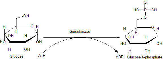
In step 1, we use the enzyme hexokinase.  The root word here is kinase.  A kinase is an enzyme that adds a phosphate group to a molecule.  In this case, hexokinase is an enzyme that adds a phosphate group to a six carbon sugar, namely glucose.  You will notice in the above picture that the enzyme is referred to as glucokinase.  Glucokinase is a specific hexokinase (Hexokinase IV), and is found in specific mammalian cells found in the intestines, liver, pancreas, and brain.

Hexokinase is generally attached to the glucose carrier found in the cell membrane.  When glucose is brought into the cell, hexokinase adds a phosphate group to the sixth carbon.  Glucose 6-P refers to a glucose molecule with a phosphate on the sixth carbon.  In order to carry out this phosphorylation, we use ATP.  ATP transfers a phosphate to the sixth carbon of glucose.  (enzymatically, how would this happen?  Would you need both ATP and Glucose in the active site?)

Why do we need to phosphorylate glucose? This is an important question, and something you should ask for every metabolic reaction.  Why do we need to do it?  What is the end product?  You should also ask yourself questions about the enzyme.

  • Why do you phosphorylate glucose?
    • Glucose is stable, so the addition of phosphate with its -2 charge causes an electrical instability in the molecule.
    • Glucose 6P is more reactive than Glucose.
    • Glucose 6P can not leave the cell through the Glucose Carrier (they are different molecules now).
    • Glucose 6P does not interfere with the concentration gradient of Glucose (they are different molecules, each with a concentration gradient).
    • Glucose then remains high on the outside of the cell, but almost zero inside of the cell (incredibly strong concentration gradient).
  • What about the enzyme?
    • Is it regulated?
      • Unidentified allosteric regulation.
    • What is the structure?
      • 465 amino acids
    • What does the active site look like?
      • Active site fits Glucose and ATP.
    • What about the activity?

Daily Challenge: Glycolysis steps 2-5
Go through steps 2-5 of glycolysis. Quote some source (book or textbook) that talks about the reaction and what is happening.  Make sure you provide the reference.  If you find a picture, add it.  After the quote, I want you to reflect on what is meant in the quote.  Most of you have not had organic chemistry yet, so I'm not asking you to delve into the chemistry behind the reaction (it is great though if you do).  What I want you to do is build a discussion of what happens in the reaction (in your own words).  Reference the quote all you like as you build your discussion.  What is important is that you build an understanding of what is happening.  Also reference the enzyme being used.  The enzyme name tells you the enzyme function.  So what is the function of the enzyme?  What reaction does it catalyze?  Is there anything interesting about the enzyme?

Thursday, February 16, 2012

Daily Newsletter February 16, 2012

BiologyMOOC Logo4

Daily Newsletter February 16, 2012

Today's Topic: Redox and Living System

Energy harvesting will be our topic next week, so I wanted to spend a moment and talk about energy in biological systems. Energy is a word that is often thrown around in various disciplines, and we define it as the capacity to do work. This definition helps to simplify a complex issue, but when it comes down to it, it really doesn't help you to understand the bigger picture of how a complex set of reactions combine to form a living system.

With ATP, you will recall, the emphasis was shifted from seeing ATP as a batter that powered reactions.  Instead, your focus was drawn to the Phosphate group, and the electrostatic effect it would have when added to a molecule or protein.  Today in lecture we saw how a phosphate could act as an allosteric regulator, turning an enzyme on or off.

With energy harvesting, I again want you to shift your focus from a nebulous form of energy.  This time, the focus will be on reducing potential.  Central metabolism describes the oxidation of glucose, so what are we harvesting?  Reducing potential.  So what is reducing potential?

A simple definition is reducing potential describes the capacity of a compound to donate electrons.  Chemistry has a strict definition involving measurements with electrodes, but for our purpose, the concept of donating electrons is what is important.

Remember the characteristics of life.  You must maintain homeostasis, and this means repair.  You have to build nucleic acids, lipids, carbohydrates and proteins.  These biosynthetic pathways often require you to reduce substrates.  To stay alive, you need a constant supply of electrons for reduction; you need reducing potential.  If you don't get these high energy electrons for reduction, you die.  We will also find that this reducing potential is needed for us to make ATP.

So, redox reactions become vital to our survival.  Redox reactions are coupled Oxidation and Reduction reactions.  One compound is oxidized as the next is reduced.
Remember, the molecules undergoing redox have to be close/touching.  But in relative size, a cell is huge compared to a simple molecule.  We may harvest electrons (oxidation) in one part of the cell, but use the harvested reducing potential in another part of the cell (reduction).  Remember, you don't have free electrons; you can't throw electrons across the cytoplasm. So, how do we couple reactions that may be separated spatially?  We use carriers!

Electron carriers, like nicotinamide adenine dinucleotide (NAD), accept electrons at the site of oxidation, and then donate electrons at the site of reduction. NAD is readily oxidized and reduced during metabolic reactions, and there is only a negligible loss of energy from the electrons carried (can we ever have NO loss of energy? why or why not?).
NAD is also classified as a coenzyme, meaning it must work with an enzyme to accept or donate electrons.  NAD can not randomly go to a molecule and oxidize or reduce it; its action is regulated by enzymes.  NAD then must bind to an enzyme that catalyzes an Oxidation, and NADH must bind to an enzyme that catalyzes a Reduction.

Daily Challenge: Action of nicotinamide adenine dinucleotide (NAD)
In the citric acid cycle is the following reaction:
In this reaction, malate is oxidized.  How do you know?  You know because NAD is reduced to NADH.  Below is a ribbon model of the protein malate dehydrogenase.  Within the protein, you will see two molecules of NAD represented as balls.  NAD binds to the enzymes active site first, and then malate binds.  Within the active site are both + and - amino acids.

Your task today, using the enzyme malate dehydrogenase, explain how enzymes work and explain how reducing potential is harvested from organic compounds.

Wednesday, February 15, 2012

Daily Newsletter February 15, 2012

BiologyMOOC Logo4

Daily Newsletter February 15, 2012

Today's Topic: Enzymes, structure and function.
  •  First thing to remember, Eynzymes are Proteins!
    • So they are constructed on ribosomes.
    • Their structure is determined by electrostatic interactions.
    • Their shape can change when things bind to it.
    • They can be denatured.
  • Second, they act as catalysts.
    • They lower activation energy by brining molecules together in the correct alignment, and can induce molecular tension to cause the reaction.
    • Though their shape may change during the chemical reaction, the enzyme is left essential unchanged at the end of the reaction.
Enzymes work by binding the substrate of the reaction, and then inducing molecular tension.  Remember, when something binds to a protein, the electrostatic interactions around the protein change, resulting in a conformational (shape) change in the protein. It is this shape change that will induce molecular tension.

Every enzyme has an active site.  This is the place where the substrate(s) will bind to the enzyme.  The active site must have a shape that loosely fits the substrate, and the electrochemical pattern of the active site must compliment the electrochemical pattern of the substrate.  When they bind, you get an Enzyme Substrate complex.  Below is a basic cartoon about the process:
Here is a quick video that shows the brief conformational change that helps to induce the reaction.


Conformational changes alone are not the only part in inducing a reaction.  You will also find that proteins can have prosthetic groups that aid them in their action.  For example, you can a Heme group with Iron that can hold Oxygen in red blood cells.  Some digestive enzymes use Chromium to help in their action.  Many metabolic pathways will contain Electron Carriers needed for redox reactions.  We will see examples of these over the next few weeks.

Daily Challenge: There are three enzymes that we will come across next week. Today, you are to look at the action of these three enzymes and articulate how they work. Use the information above as guidance, but be specific for each of these three enzymes. One point to look at is whether or not these enzymes require additional prosthetic groups or coenyzmes/cofactors. The enzymes are glucokinase (hexokinase 4), glyceraldehyde-3-phosphate dehydrogenase, and aldolase. [Wikipedia warning: be careful with wikipedia on this one. Some enzymes have good descriptions, while others are either too technical or poorly written.]

Admin Note: As of today, only 7% of the students have started the calibrations. Do not delay. You will only hurt yourself if you wait until the last minute.

Tuesday, February 14, 2012

Daily Newsletter February 14, 2012

BiologyMOOC Logo4

Daily Newsletter February 14, 2012

Administrative Note: CPR Calibrations
Make sure you start your calibrations today. Do not wait until the last day to do all of calibrations and reviews. Give yourself time to do them. Pace yourself by doing one task each day until the close of the assignment. I have given you til Sunday to finish this task.

Daily Topic: Adenosine Triphosphate
You have most likely heard ATP referred to as the "energy currency" of the cell. In fact, your textbook uses this analogy: "Just as it is more effective, efficient, and convenient for you to trade money for a lunch than to trade your actual labor, it is useful for cells to have a single currency for transferring energy between different reactions and cell processes." This is a lovely fiction that does not serve molecular biologists. It is a convenient expression, but it conveys a very serious misconception.

Basically put, nucleotide triphosphates have their foundation in the nucleotide structure, with the addition of extra phosphate groups. Adenosine Triphosphate is the most prevalent nucleotide triphosphate. The picture below show the general structure of ATP.

You have three phosphate groups, each with a negative charge, covalently bonded to each other.  The phosphate groups naturally want to repel each other, but they are held together by one of the strongest bond types.  What does this mean?  Molecular tension!  But it must be noted that ATP is stable.  It does not spontaneously loose phosphates (if it did, you would also release heat).  It takes enzymatic action to remove the phosphate. When a phosphate is removed from ATP, it is generally attached to another molecular structure (enzymes, sugars, etc...).  The exception to this will be in building nucleic acids.

So what is the misconception with "energy currency"?  A better question will be to ask, what is energy to a cell?  Cells use reducing potential for energy; electrons harvested from reduced compounds provide reducing potential.  This is needed in order to make many biochemicals including ATP.  What then is ATP?

The concept of ATP as an "energy currency" comes from ATP turning on enzymes or assisting an enzyme during a "power step" in a metabolic pathway.  But ATP does not add energy; it just rearranges charge distribution around a molecule (electrochemistry).  Remember, the phosphate group is negatively charged (-2). 
When you add a phosphate group to a protein, you change the electrical signature around that portion of the protein (same will be true of other molecules as well).  What will happen to the protein?  It will change shape (conformational change).

As we will see in upcoming weeks, this change of shape is critical to enzyme function.  You have already encountered this once before, with the Sodium/Potassium ATPase.

Daily Challenge: Function of ATP
Today, I want you to discuss the function of ATP. Do not describe it as an energy currency, instead describe how the addition of phosphates cause a change in the electrochemistry of a protein, and how that affects the conformation of the protein. Use Myosin in muscle cells as your example. We have not covered Myosin, but it is a very easy model for how ATP acts.

Monday, February 13, 2012

Daily Newsletter February 13, 2012

BiologyMOOC Logo4

Daily Newsletter February 13, 2012

Daily Topic: Thermodynamics and Equilibrium

Today you are asked to reflect upon the topics of thermodynamics and equilibrium as they pertain to biology. These are topics originally introduced in chemistry, so you may want to go back to your chemistry books to refresh your memory. While biochemists may use these concepts unaltered from their original meaning in chemistry, most biologists look at these two concepts from a slightly different perspective.

The Laws of Theromodynamics: (the two important ones for biology are in bold).
0. If two systems are in thermal equilibrium with a third system, they must be in thermal equilibrium with each other. (Kind of a no brainer, but it is something that comes up in biology).
1. Energy can neither be created nor destroyed, but can be changed from one form to another.
2. In all energy exchanges, if no energy enters or leaves the system, the potential energy of the state will always be less than that of the initial state.
3. All processes cease as temperature approaches absolute zero. (not generally a concern to biologists).

Law 1 and 2 are the ones we most often use in biology, but how?
The first law of thermodynamics is rather simple. We convert one type of energy into another.
  • Phototrophs are organisms that can convert light energy to chemical energy.
  • Motile organisms (such as animals) can convert chemical energy into mechanical energy.
  • Luminescent organisms can convert chemical energy into light energy.
  • All organisms can convert one type of chemical energy into a different type of chemical energy.
Later in the week, we will discuss redox reactions, but for now I want to put this concept into your heads:  One of the most critical energy exchanges in biology will be the production of reducing potential.

The second law plays a role in homeostasis.  With the second law, we know that energetically, we can never break even.  Every time that we undergo a chemical reaction, we loose energy.  This loss is generally going to be as heat, which is also not good for a cell (too much heat, and the cell boils).  So we have be as efficient as possible knowing that we are always loosing energy.  How does this play out?
  • Plants take in sunlight, and make glucose.
    • The sunlight is high energy.
    • The glucose is going to have to have less energy because we used photons to excite electrons (an energy change).
  • Animals eat plants.
    • We catabolize the glucose for energy (reducing potential).
    • Do we get 100% of the energy in glucose?  NO.
  • Animals build biomass (make proteins, lipids, etc... as needed for life).
    • The energy we got from glucose is further lost when we make new biochemicals.
    • We constantly need energy to keep rebuilding ourselves.
    • We constantly need energy inputs to maintain homeostasis.
How does equilibrium play a role?  This come in how we experience metabolism.  In chemistry lab, to do multiple reactions, you first converted A èB, then you took B èC.  You may have moved them to different test tubes.  If nothing else, you waited until the first reaction reached equilibrium before proceeding to the next reaction.  In other words, you waited for A to convert to B, then B to C.  In living systems, there is a near seamless transition between reactions.  The product of one reaction almost instantaneously becomes the substrate for the next reaction.  A misconception is that cells never reach an equilibrium, but this is not true.  Cells are always in a state of dynamic equilibrium. 


Daily Challenge:
Today you are tasked with describing in your own words how the laws of thermodynamics and eqiulibrium play out in living systems. Use the above a a jumping off point, but build your own discussion, examples or analogies.


Special Blog Opportunity: You can earn up to 4 points by reflecting on Milestone Exam 1. Go over the text, and reflect on each of the questions you got wrong. To earn points, you must perform the following:
1. Explain why the keyed answer was the correct answer for any question you missed.
2. Provide a reference for the correct answer.
3. Explain why you got the question wrong.

Sunday, February 12, 2012

Weekly Update - Week 6 - Enzymes and Energetics

BiologyMOOC Logo4

Weekly Update Week 6 - Enzymes and Energetics


Administrative Note: CPR
Your paper is to be uploaded by 11:55 pm. Please note you will have to finish the pre-test and tutorial before you can access the assignment. DO NOT WAIT UNTIL THE LAST MINUTE!

When you first log into the site, you will use your Panther ID.
Do not use the initial zeros of your panther ID! If you keep the initial zeros, the system will say that you are not registered. Just put your Panther ID in again, this time without the zeros.

At present, the system is preventing you from doing the calibrations until the test submission time period is over.  You will be able to perform calibrations starting tomorrow.


Weekly topic: Enzymes and Energetics.
This week we move into metabolism. Our first stop will be a review of thermodynamics, an introduction to enzyme kinetics, a look at ATP, and finally an introduction to enzymatics.


Learning Objectives:
  • Be able to discuss the laws of thermodynamics as they relate to living systems.
  • Be able to discuss chemical equilibrium as it relates to living systems.
  • Be able to discuss the structure and function of ATP.
  • Be able to disucss how enzymes, as catalytic agents, speed up reactions.
  • Be able to describe how enzymes work.
  • Be able to discuss the regulation of enzyme activity.
  • Be able to discuss the importance of redox reactions to living systems.

Friday, February 10, 2012

Daily Newsletter February 10, 2012

BiologyMOOC Logo4

Daily Newsletter February 10, 2012


Today's Topic: Evolution Friday

One of the characteristics of life is that cells must have the ability to sense and respond to their environment. Cell communication is part of this ability, but how did it come about? Reflect upon this question.


Daily Challenge:
Consider the question above. You will not find one source that discusses the evolution of signal transduction, but with what you know of evolution, begin to form a hypothesis about how chemical communication and signal transduction may have started, and how it became more complex. Understanding how signal systems evolve can give insight into a number of complex issue from neural circuity to hormone imbalance.

Thursday, February 9, 2012

Daily Newsletter February 9, 2012

BiologyMOOC Logo4

Daily Newsletter February 9, 2012

Administrative Notes:
Due to a technical error, CPR has not opened the assignment. It will be opened later today. An announcement will be sent out when it is open. Until then, if your paper is ready, make sure you take some time to review and edit before uploading.


Today's Topic: Protein Kinases

Kinase refers to a group of enzymes that transfers a phosphate group from one molecule to another. The term applied to this process is Phosphorylation. Notice that the word kinase ends with the suffix -ase. This suffix is used to denote an enzyme, and you will see nearly every enzyme ending with the suffix -ase (remember this). Protein Kinases refer to a subgroup of kinases that phosphorylate proteins.

Kinases are generally named for the substrate that they phosphorylate. So, myosin heavy chain kinase will phosphorylate the heavy myosin chain. Cdk-activating kinase will phosphorylate another enzyme known as CDK (CDK is important in regulating cell division, and we will come back to this enzyme in a few weeks).

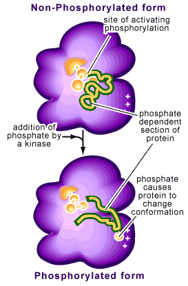
Remember that enzymes are proteins folded into at least a tertiary structure, and that protein structure is determined by electrochemistry of the individual amino acids. A phosphate carries a -2 charge. When you bond a phosphate group to an enzyme, you alter the charges of the molecule, and thus alter the shape of the molecule. This shape alteration is generally referred to as either a confrontational shift or conformational shift (the latter term is more common). This shape alteration will result in either the activation or deactivation of the protein/enzyme. The following cartoon will give you a good visual of the effects of phosphorylation.


Daily Challenge:
In your own words, describe how the action of a protein kinase can alter the function of a protein. Use the Insulin Signal Transduction pathway as your means of discussing kinase activity.

Wednesday, February 8, 2012

Daily Newsletter February 8

BiologyMOOC Logo4

Daily Newsletter, February 8, 2012


Administrative Notes:  Milestone Paper 1


Starting tonight, you will be able to upload your milestone paper.  The upload period will continue until Saturday evening. You will be notified if there is any change to this schedule.


Today's Topic:  Indirect Transduction
Many of our signals work through a process of indirect transduction, which means that the initial signal (primary signal) is converted into a secondary signal that causes the ultimate cellular effects.  One of the most well studied indirect systems is seen in the use of a G-Protein linked receptors.   


Daily Challenge:  G-Protein as a mechanism of indirect transduction.
Review material in your textbook about indirect signal transduction.  Explain the function of  the G-Protein by using the formation of the IP3 and DAG secondary messengers.

Monday, February 6, 2012

Daily Newsletter February 6, 2012

BiologyMOOC Logo4

Daily Newsletter, February 6, 2012


Administrative Notes:  Milestone Exam 1
Tonight at 5pm, you will be able to start your first milestone exam.  The exam link will be located on the uLearn course home page.  The exam will end on Tuesday at 5pm.  You will have 50 minutes for the exam, and only one attempt.  I strongly encourage all students to take the exam tonight.  If there is a technical difficulty, please contact me ASAP at rmaxwell@gsu.edu.

Do not wait until the last minute!  If you wait, I may not be able to help you if there is a technical problem.  Remember, the exam is only open for 24 hours and will not reopen.  There are no make-ups.


Today's Topic:  The basics of cell communication
This week marks our discussion of cell to cell communication.  The first thing which must be remembered is that cells must be able to sense their environment, other cells, and process information from other cells.  Signal reception occurs in all known organisms (from picking up environmental to cellular signals).  For instance, in bacteria, we know of a signal phenomena known as Quorum Sensing.  With this signal system, bacteria release chemicals as they grow which other individuals of the same species (and some times other species) pick up; when the concentration of the chemical reaches a critical point, cellular changes can be observed in the community.  Basically, as the population increases, cells begin to change (we will talk about this more later in the week).

But how do cells pick up signals?  Signal recognition begins with protein receptors.  For a cell to pick up, or register, a signal, it must build a receptor for that signal.  This sets up another question:  what is a signal?

Most of the signals we will talk about are chemical signals, meaning we have a chemical compound that will "fit" a receptor, activating it.  There are other signals though:  light can be a signal (photoreception), temperature (thermoreception), and even mechanical such as touch (mechanoreception).  As mentioned, out discussions for this week will focus on chemical signal pathways.

By far, the most common type of signal system will involve chemicals.  Hormones are chemical signals, neurotransmitters are chemical signals, even carbon dioxide is used as chemical signal in the human body.  Because there are so many different chemical signals, we have a generic word for any compound that could bind and activate a receptor protein:  Ligand.  As a general word, ligand is used when we discuss the basic concepts of signal systems.

At their most basic, a chemical signal system (or pathway) will be comprised of a Ligand and a Receptor.  When a ligand binds to a receptor (ligand-receptor complex), the receptor changes shape (conformation), which elicits a physiological response in the cell.  Remember:  The receptor is a protein, and when proteins change shape, they have an effect on the cell.  So the basic signal system will be:  Ligand binds to receptor, receptor changes shape, cellular response occurs.


Words of the Day:  Paracrine & Autocrine
Prepare definitions for these two words and put them in your notes.

Daily Challenge:  A Model Signal Transduction Pathway
Review the model signal transduction pathway of the ompC gene in Escherichia coli as discussed in the Sadava textbook.  Write a general description of how the signal/receptor complex alters cell physiology.  Describe the nature of the ligand and the receptor.

Friday, February 3, 2012

Weekly Update 5 - Cell Communication

BiologyMOOC Logo4

Weekly Update 5 - Cell Communications

This week we discuss the basics of cell communication. We have also arrived at your first Milestone, so your time management skills will be tested. On Monday, February 6th, at 5pm, your Milestone exam will open. You will have one attempt on this exam, and it closes at 5pm on February 7th. On February 8th, you can start uploading your Milestone Papers to CPR. During the week, I will also be sending newsletters and challenges. So pick your battles this week.

Admin note: On the calender, it says we will meet on Wednesday February 8th for lecture. This is an error. We will only meet this Thursday, February 9th, for lecture.


Suggested Readings:
You are asked to review chapter 7, Cell Signaling and Communication, from the Sadava text. What does it mean to review a chapter? Take 30 minutes to skim the chapter, then use it as a reference when it comes to challenges. Do not try to commit the chapter to memory, just review it.


Learning Outcomes:
  • Be able to describe what is meant by cell communication.
  • Be able to describe the basic concepts/steps in a signal transduction pathway.
  • Be able to describe the general function and classification of receptors.
  • Be able to describe the basics of various transduction models.
  • Be able to describe the G Protein-cAMP model in more detail.
  • Be able to discuss the basics of how cells respond to signals.
  • Be able to discuss direct cell-cell communication.

Daily Newsletter February 3, 2012

 
 Daily Newsletter                                        February 3, 2012


Today's Topic: Evolution Friday

At the end of the cell structure chapter in the Sadava text is a section on the evolution and origin of organelles.  Beyond theory, one way to study the development of cellular structures or specializations is to look at existent organisms that do not fit the typical cellular model that we reviewed this week.  These anomalous structures can give us insight into possible evolutionary steps. 


Daily Challenge:
Review the section of your text on the origin of organelles.  What follows is a list of organisms or terms that describe some type of anomaly in typical cell models.  Pick one to write about.
  • Zooxanthella
  • Planctomycetes
  • Borrelia burgdorferi
  • Rhodobacter 
  • Amitochondrial

Thursday, February 2, 2012

Daily Newsletter February 2, 2012

 
 Daily Newsletter                                        February 2, 2012


Today's Topic: The Cytoskeleton

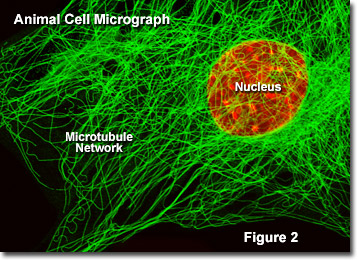
The cell is not just a fluid filled sack, but an organized structure complete with internal supports.  Consider it a highrise building that is just squishy.  Creating the structure are a protein fibers (structural proteins).  Structural proteins typically are in quatrenary structures, meaning that individual proteins combine to create the overall structure.  The cytoskeleton is composed of Microtubules, Microfilaments, and a group of proteins referred to as the Intermediate Filaments.

The microtubules are one of the most well studied cytoskeletal elements.  They are composed of the protein tublin, and are responsible for the flagella, mitotic spindles, internal structure, and movement of materials and vesicles within the cell (they act as roads on which vesicles are carried). 

Microfilaments are composed of actin.  They help to form structure, and are used in movement, such as mucle contraction, the formation of pseudopods and bulk transport. 

The intermediate filaments are a family of structural proteins that are smaller than microtubules, but larger than microfilaments.  There are a number of different types, and some are specific for a given cell type.  Wikiepdia provides a good list of the different types of intermediate filaments in the article itermediate filaments.

Spectrin is a structural protien that was originally classified as an intermediate filament.  In recent years, the importance of this protein to the cell membrane has become more noted, so it deserves it's own place.  The structure of spectrin has it reclassifed as related to actin (microfilaments).


Daily Challenge:  Cytoskeleton
Today, you are to describe in brief the function of the cytoskeletal elements listed above.  You do not need to go in depth about the structure, at present the function is more important.  Pick one of the above cytoskeletal elements to be your focus today.  Write more about that element, including it's structure.

Wednesday, February 1, 2012

Daily Newsletter February 1, 2012

 
 Daily Newsletter                                        February 1, 2012


Today's Topic: The Mitochondria

The mitochondria is known as the power house of the cell, for this is where eukaryotic cells experience oxidative phosphorylation and ATP production.  We will come back to ATP in a latter newletter, but you should note that phosphorylation of proteins is a powerful activator of enzymes (allowing them to work).  Everything from pump systems, cellular movement, and even muscle contraction relies on ATP.

Like the structure of the nuclear envelope, the mitochondria is a double membrane bound structure, but the origin of the nuclear envelope and mitochondria are very different.  It is hypothesized that the nuclear envelope formed from the infolding (invagination) of the cell membrane.  The mitochondria, in contrast, is two separate and distinct membranes.


The Endosymbiotic Theory is used to explain the development of the mitochondria.  (Question: why should we consider this a theory?)  Before we get to the endosymbiotic theory, we need to first look at the structure of the mitochondria:


We have an outer membrane and an inner membrane.  Between the two membranes is the Intermembranous Space.  The inner membrane is highly folded into Cristae (Question:  why would you fold a membrane?).  The inner compartment, bounded by the inner membrane, is known as the mitochondrial matrix.


With the structure in mind, how does this differ from the nuclear envelope?  
  • The outer membrane displays eukaryotic proteins.
  • The inner membrane displays prokaryotic proteins.
  • The intermembranous space stores hydrogen ions, so is acidic.
  • The matrix contains a circular bacterial DNA molecule and 70s (prokaryotic) ribosomes.
  • The mitochondria is self-replicating (the DNA can make copies).
 The endosymbiotic theory describes the mitochondria as a bacterial symbiont that was engulfed by a "proto-eukaryotic" cell.  A relationship formed between the two cells, with the mitochondria taking over ATP production, and the "proto-eukaryotic" cell loosing the ability.


Genenomic analysis of the mitochondria shows that it comes from the bacterial Order  Rickettsiales, which means that it is related to the intracellular parasite Rickettsia rickettsii (Rocky Mountain Spotted Fever).  Mitochondrial genes are inherited matrilineally, and are the basis of human population genetics studies of the mitochondrial genome.


Daily Challenge:
Write about the mitochondria, the endosymbiotic hypothesis and human mitochondrial genetics.  Explore these topics, and feel free to go deeper on any feature of the mitochondria that interests you.  One question I want you to focus on is why is the mitochondrial genome reduced (smaller) than other members of the Rickettsiales?