Thursday, February 20, 2014

Microbiology Daily Newsletter February 20, 2014 - Transcription Termination

Site LogoMicrobiology Daily Newsletter

February 20, 2014 - Transcription Termination


Transcription Termination

 The elongation phase of transcription (polymerization of RNA) occurs the same in all domains, working via base complementarity.  Termination, like initiation, is different.  In bacteria, transcription termination occurs either through an intrinsic termination system (Rho-Independent) or through the use of a Rho (ρ) factor (Rho-Dependent).

The Intrinsic System (Rho-Independent) 

After a the coding region of the gene, or the final coding region of an operon, there will be an inverted repeat of nucleotides on the DNA.  Following this repeat will be a series of six adenine nucleotides.  When this region is translated, the inverted repeat (usually rich in G-C) will anneal, creating a hairpin loop structure.  The six adenine nuclotides are transcribed into uracil.

A protein associated with the RNA polymerase, nusA, catches the hairpin loop structure, and holds it.  This stalls transcription in the region where Uracil has been transcribed.  A region of A-U in a DNA-RNA duplex (i.e., strand of RNA bound to a complementary DNA strand) is very weak (it is a major weak point).  This naturally dissociates from the DNA strand, and is released by the RNA polymerase.

Summary:  The intrinsic system relies on the stalling of a hairpin loop at the end of the transcript, and a region of A-U, to terminate transcription.


The Rho-Dependent System

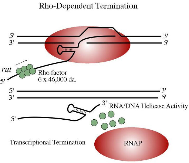
The ρ-Factor is an ATP-dependent hexameric helicase, and binds to a cytosine rich region of RNA >70 bases upstream from the termination site.  This cytosine dominant region is known as the Rho Utilization Site (abbreviated rut).  Once bound, the ρ-Factor moves up the RNA strand toward the 3' end (e.g., where the DNA-RNA duplex is found).  RNA Polymerase pauses at the termination site, and during this time, the ρ-Factor comes catches up to the transcription fork (DNA-RNA duplex), and unwinds it.  Transcription ends.

Comparison between the two termination systems
Comparision of intrinsic termination and Rho-mediated termination.  Greive S.J. and P.H. von Hippel. (2005) Thinking quantitatively about transcriptional regulation. Nat Rev Mol Cell Biol 6:221-32.


Eukaryotic Transcription

To refresh your memory regarding Eukaryotic transcription, you may want to read over these articles:

Daily Challenge 

Your goal today is to discuss the differences similarities and differences between bacterial and eukaryotic transcription, specifically initiation and termination.  Read DNA Transcription in case you need a refresher.
REMEMBER to discuss both similarities and differences.  While doing this, consider evolutionary differences.  

Wednesday, February 19, 2014

Microbiology Daily Newsletter February 18, 2014 - Transcription Initiation

Site LogoMicrobiology Daily Newsletter

February 18, 2014 - Transcription Initiation


Hopefully you remember the basic idea of a gene and the mechanism of transcription as it applies to eukaryotic cells.

For bacterial transcription, we will first look at the promoter.  You may recall the TATA box with eukaryotic promoters.  In bacteria, we find the Pribnow-Schaller box, commonly just referred to as the Pribnow box.  The Pribnow-Schaller box is a six nucleotide sequence TATAAT. QUESTION:  Why six nucleotides?

With eukaryotic promoters, you may recall that the TATA box is not the only recognition sequence needed.  This is also true of prokaryotic promoters.  The Pribnow-Schaller box is found at -10 from the start of the gene.  The complete promoter also contains a -35 recognition sequence, and is also comprised of six nucleotides.  The -35 promoter element usually has a sequence of TTGACA, but note that this can vary among different bacterial taxa (usually at the class or family level taxa).  NOTE:  The -10/-35 promoter is used for normal house keeping genes.  We will see some variation shortly.

The prokaryotic RNA Polymerase catalyzes the reaction of both coding and non-coding RNA, unlike eukaryotes that have job specific RNA polymerases (for example, you may remember that RNA-Pol I forms the 45S pre-rRNA, while RNA-Pol III forms tRNAs).  The prokaryote RNA Polymerase Complex is a holoenzyme composed of RNA Polymerase and a Sigma (σ)factor.  The eukaryotic RNA Polymerase complex has a specific RNA polymerase and a variety of initiation factors.

Below is an example of the bacterial transcription initiation complex.  Note that you are seeing RNA Polymerase with 2 different σ factors, and that the different sigma factors have slightly different promoter recognition sites.  When they bind, they move from a closed complex to an open complex; the open complex gaining its name from the opening of the DNA helix.
Add cFrom: Bush M , and Dixon R Microbiol. Mol. Biol. Rev. 2012;76:497-529.  Initiation of transcription by the RNAP-σ70 (A) and RNAP-σ54 (B) holoenzymes. The σ70 factor directs the binding of polymerase to the consensus −10 (TATAAT) and −35 (TTGACA) sequences to form an energetically unfavorable closed complex (CC) that is readily converted into an open complex (OC) to initiate transcription. In contrast, the σ54 factor directs the binding of RNAP to conserved −12 (TGC) and −24 (GG) promoter elements that are part of the wider consensus sequence YTGGCACGrNNNTTGCW (where uppercase type indicates highly conserved residues, lowercase type indicates weakly conserved residues, N is nonconserved, Y is pyrimidines, R is purines, and W is A or T) (10). This forms an energetically favorable CC that rarely isomerizes into the OC. In order to form the transcription “bubble,” a specialized activator (a bacterial enhancer binding protein [bEBP]) must bind and use the energy from ATP hydrolysis to remodel the holoenzyme. aption
Once the RNA Polymerase-σ factor Holoenzyme is bound in the open complex form, transcription can proceed to the elongation phase.  The next issue specific for prokaryotic transcription will be termination.


Daily Challenge

Sigma factors play a critical role in coordinating bacterial cell physiological states.  Below is a list of common sigma factors.


Sigma Factor
Gene
Function
s70
RpoD
Primary s factor, Housekeeping
s19
FecI
Regulates fec gene for iron transport
s24
RpoE
Extreme heat stress
s28
RpoF
Flagellar genes
s32
RpoH
Heat shock
s38
RpoS
Starvation/Stationary phase

Along with these can be found anti-sigma factors that block the function of expressed sigma factors (a form of regulation).  Discuss the role of sigma factors as means of global gene regulation.  Why is having a sigma factor system beneficial to bacteria?  How does this differ from eukaryotic promoters?

Monday, February 17, 2014

Microbiology Daily Newsletter February 17, 2014 - Bacterial Genetics

Site LogoMicrobiology Daily Newsletter

February 17, 2014 - Bacterial Genetics


At this point, all students should be able to provide a solid description of Replication, Transcription and Translation.  Therefore, we will not concern ourselves with the basics of these processes, but instead focus on how Bacteria differ from Eukarya in these three processes.  As we move though this week, we will also begin our discussion on genetic regulations.

The following topics in genetics you should have a strong familiarity with.  If you don't then it is time to brush up on your genetics.

  1. Be able to discuss replication, transcription and translation.
  2. Be able to describe in detail the mechanisms of replication, transcription and translation.
  3. Be able to discuss the concept of a genome, and what constitutes a genome.
  4. Be able to discuss supercoiling of DNA and the use of topoisomerases.
  5. Be able to discuss how DNA analysis is done, and why.
  6. Be able to discuss how researchers analyze the whole genome, and why it is important.
  7. Be able to discuss all of the different types of RNA.
  8. Be able to discuss post-translational modification of proteins.
  9. Be able to describe open reading frames, paralogs, orthologs and DNA alignments.
  10. Be able to discuss various mechanisms of gene regulation and epigenetics.

 Bacterial DNA Replication

 The genetic process of replication is fundamental to all living systems, and the basic mechanism is the same: DNA is unwound and opened, each parental strand acts as a template upon which a newly synthesized strand is based; the resulting daughter molecules are semi-conservative, being formed of one template (old) strand and one new strand.  You should be intimately familiar with this process.

So, what is different between bacterial replication and eukaryotic replication?
Remember that the nucleoid (genophore) of the bacteria is a single circular molecule that is vastly smaller than eukaryotic chromosomes.  Unlike the chromosome, which can have multiple origins of replication, bacteria will generally only have 1 origin of replication (Ori).

Eukaryotic chromosomes are linear, and so you keep replicating til you get to the end (remember, there is a problem with the ends potentially degrading...remember telomerase?).  Bacterial DNA is circular, so there is no end.  Instead, you find a terminus, or termination point (Ter).

As with all DNA replication, you will get two replication forks on either side of the origin of replication, these will then meet at the terminus.  All the while, the DNA is anchored to the cell membrane, and it is the movement of the cell membrane that will separate the two daughter molecules.

The process begins when DnaA binds to the Ori.  DnaA, and the specific sequence at Ori, are genera/species specific.  The most highly studied system is Escherichia coli, where a repeat of 5' - TTATCCACA - 3' is used as the binding sequence for DnaA.

DnaA enzymes needed for replication including Helicase,  DnaB, and DnaC.  DnaB anchors at what will become replication forks, and aids in the binding of primase.

Single-stranded binding proteins and DNA gyrase will be needed to prevent re-annealing and positive supercoiling respectively.  DNA polymerase III holozyme will be the primary work horse of the elongation phase.

Termination is interesting, as the two replication forks are forced to meet in the Ter region, by a process referred to as the replication fork trap.  The two daughter strands are joined as interlocking rings in the terminus (catenane, or mechanically linked molecular architecture).  DNA Topoisomerase IV will be the critical player in unlinking the daughter molecules, and will need DNA gyrase to assist.
From: Charvin G et al. PNAS 2003;100:9820-9825.  Configurations of a circular replicating DNA. (a) Model for in vivo strand separation: the progression of the replication complex leads to the formation of (+) supercoils (L-nodes) in front of the replication fork and R-precatenanes behind. Topo IV removes (+) supercoils and gyrase generates (–) supercoils so that, under the action of both enzymes, the chirality of the precatenanes is inverted. Topo IV may then unlink the molecules by removing the L-catenanes. (b) Model for in vitro decatenation by Topo IV: R-catenated plasmids may form L-supercoils that are removed by Topo IV with a high rate [as observed in our experiments and some bulk assays (21)]. However, the removal of the last few links is slow , because Topo IV will relax only a rare fluctuation of an R-node to an obtuse angle that is similar to its preferred substrate: an L-node with acute angle (see Fig. 1b).


Daily Challenge

Review DNA replication as it pertains to bacteria, and consider, how would you move from the replication of a circular DNA molecule to a linear molecule?  What would need to change?  Initiation? Elongation? Termination?  How different are each of these process from eukaryotic replication?

Tuesday, February 4, 2014

BOLO Microbiology Daily Newsletter February 4, 2014 - Metabolism & Growth

Site LogoMicrobiology Daily Newsletter

February 4, 2014 - Metabolism & Growth


Metabolism and Growth are both essential characteristics of life, and are intimately related to each other.  You may recall that living systems require CHONPS (carbon, hydrogen, oxygen, nitrogen, phosphorous, and sulfur) to make biomolecules.  In addition, we need minerals, trace minerals, and energy in the form of reducing potential (i.e., electrons).  The acquisition of carbon and energy are the primary focus of central metabolism (glycolysis, citric acid cycle, etc...) and photosynthesis.  But what about the other needs?

Amino acids and nucleic acids both need nitrogen.  Nucleic acids need phosphorus in the form of phosphate groups.  Sulfur is needed to form disulfide bridges and a variety of thiols.  Phosphates become a prime regulator of protein function, and ions are needed to establish membrane potentials.  All of these are needed to increase the biomass (growth) of cells.

In looking at microorganisms you need to become accustomed to the metabolic needs and diversity of cells.  Though less than 1% of bacteria are culturable, these organisms have helped us understand the dynamic relationship between nutrition, metabolism and growth.  They have also provided researchers with known genes that can be used to understand potential functions from genetic analysis.

Today, your goal is to understand how variations in growth conditions can affect bacterial growth, both in terms of biomass accumulation and increases in population size (binary fission).


Heterotrophy vs Autotrophy 

These two terms describe the acquisition of carbon. Heterotrophy is used to describe organisms that rely on organic carbon as building block. Autotrophy describes organisms that have the ability to fix carbon dioxide into an organic compound (carbon fixation means adding a CO2 onto an existing organic carbon backbone; this makes the carbon biologically available).

Phototrophy vs Chemotrophy

These two terms describe the acquisition of energy in the form of reducing power.  Phototrophs have the ability to utilize photons to generate high energy electrons that can be used as reducing potential.  Chemotrophs utilize reduced compounds for reducing potential (i.e., energy).

The concept of chemotrophs can be further divided in to chemoorganotrophy, in which cells utilize reduced organic compounds for energy, and  chemolithotrophy, where cells utilize reduced inorganic compounds for energy.

Nitrogen

Molecular nitrogen is the most abundant gas in our atmosphere, but it is not very accessible by most biological organisms.  For nitrogen to become available, it needs to be reduced.  Atmospherically this is accomplished by electrical activity, but far more becomes available through biological means.  Nitrogen fixation is an important chemical pathway, and is the first step in the nitrogen cycle (the movement of nitrogen through ecosystems).  The diagram below provides an explanation of nitrogen fixation:
Plant Biotechnology Handbook by Niir Board.
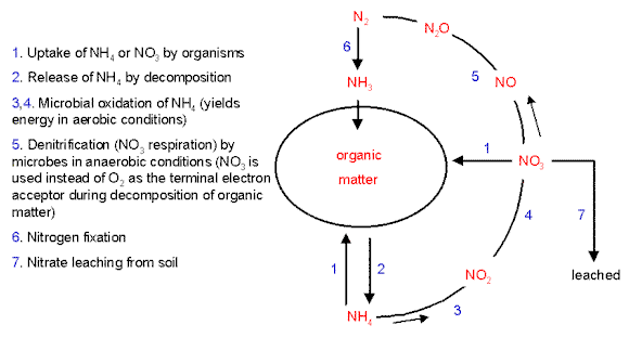
Note the amount of redox reactions needed, and the requirement for Mo (Molybdenum) as a cofactor for the enzyme nitrogenase (looking at the nitrogenase cycle, how is molybdenum used?).  Notice also that Magnesium (Mg) is also needed during the reduction of molecular nitrogen.  Nitrogen has a number of biologically active states:  including NH3, NH4, NO3, NO2, and NO.  The movement of nitrogen through living systems is shown in the diagram below:
Nitrogen fixation.http://archive.bio.ed.ac.uk/jdeacon/microbes/nitrogen.htm
Can humans use raw NH3?  How about bacteria?  Can all bacteria use NH3, or only some?  These questions come to the heart of today's topic.  All bacteria need nitrogen, but they differ in how they acquire nitrogen.  Some can only harvest nitrogen from organic compounds, while others can make use of some of these other compounds.  Bacteria also have to give off nitrogen as waste, but how do they do it?  We convert ammonia to urea, but do bacteria need to make urea?  This also brings up another point about living in a community of different types of bacteria:  the waste of one cell, may be the food for another cell.


Daily Challenge

What determines the metabolic diversity of a bacterial species?  What other conditions do you need to consider when growing bacteria?  How does central metabolism provide raw materials needed for growth?

Monday, February 3, 2014

BOLO Microbiology Daily Newsletter February 3, 2014 - Biosynthesis

Site LogoMicrobiology Daily Newsletter

February 3, 2014 - Biosynthesis


The flip side of the catabolic pathways are the anabolic pathways.  For cells to grow, they must produce biomass: proteins, lipids, and in the case of bacteria, peptidoglycan.  Up until now, the focus of most biology classes is on the catabolic processes, with an enphasis on energy harvesting.  It is important to remember that the catabolic processes also provide carbon building blocks as well as energy in the form of reducing potential.  The cell must balance a need for energy with a need for carbon building blocks. Today, we are going to look at two biosynthetic (anabolic) pathways: amino acid synthesis and Polyhydroxyalkanoates (PHA) synthsis.  

α-ketogluterate family: Glutamine Biosynthesis

Glutamine is an amino acid derived from α-ketogluterate.
 We begin with the citric acid cycle intermediate α-ketogluterate, then by adding an amino group, we produce the amino acid glutamate.  Glutamate can then converted to glutamine by adding a second amino group.  NOTE that the addition of a free amino group requires a phosphorylation, and then a substitution with the phosphate.  Why do you think this is required?


Oxaloacetate/Aspartate Family of Amino Acids


The citric acid cycle intermediate oxaloacetate can be used to make a wide range of amino acids.  The first reaction is a transamination in which an amino group from an existing amino acid is transferred to oxaloacetate to produce the amino acid aspartate.  Aspartate, as can be seen, can be used to produce isoleucine, methionine and leucine.  Notice that the production of lysine will require substrate reduction using NADPH and the addition of a pyruvate.  

Question:  How is NADPH different from NADP?  Why does a cell have two different electron carriers based on nicotinamide adenine dinucleotide?


Polyhydroxyalkanoates (PHA) Biosynthesis

 Polyhydroxyalkanoates (PHA) is a large carbon polymer produced by some bacteria as an energy storage inclusion body. To the left is
Poly-(R)-3-hydroxybutyrat, and example of the PHA group.  The monomers are attached via an ester bond, making this a biologically produced polyester.  It is a plastic, and a common example of a bio-plastic, such as you might find used in grocery store plastic bags.
PHA pathway based upon Verlindin RAJ, Hill DJ, Kenward MA, Williams CD, and I Radecka. 2007. Bacterial synthesis of biodegradable Polyhydroxyalkanoates. Journal of Applied Microbiology 102:1437–1449
The above pathway demonstrate the production of 3-hydroxyacyl-CoA, and the the polymerization into PHA.  Remember that this is a process that allows bacteria, such as Bacillus sp. and Rhodococccus sp. to have an energy storage solution.  The compound though is also effective industrially.


Daily Challenge:

Examine the balance cells must maintain between energy and building blocks by looking at cellular requirements for biosynthesis.  Remember that before divisions, cells must grow in biomass.  Use as your example a biosynthetic pathway shown in your book or that you find online (do not use the ones provided).  You can use amino acid, lipid or nucleic acid production for example, or you can look at some of the other products.  A reference you may find useful is GLAMM: Genome Linked Application for Metabolic Maps.  GLAMM provides an interactive map of known metabolic pathways with genomic links. The map can be limited to various microorganisms that you specify.