Tuesday, January 28, 2014

BOLO Microbiology Daily Newsletter January 28, 2014 - Metabolic Diversity

Site LogoMicrobiology Daily Newsletter

January 28, 2014 - Metabolic Diversity


Prokaryotes have amazing metabolic diversity, unlike eukaryotes which are metabolically very similar. Bacteria and Archaea can utilize an incredibly diverse pool of carbon, nitrogen, phosphorus and sulfur compounds. Individuals have been shown to use unusual metals, produce compounds never thought to exist naturally, and even degrade compounds we believed were refractory (or non-biodegradable).

For example, 2,4,6-Trinitrotoluene (TNT) can be degraded by Pseudomonas spp.  Below is a diagram showing proposed and investigated degradation pathways.
Esteve-Núñez A et al. Microbiol. Mol. Biol. Rev. 2001;65:335-352
Do you find a metabolic diagram like this confusing or intimidating?  Take a deep breath, and calmly look at the diagram.  In the center you see TNT, our starting compound.  From there you see arrows going to different products.  Some of the arrows have numbers; these represent citations in the review article where this image was published.  The citations are showing you where the original work on this pathway can be found.

From here, think about what you know of metabolism and organic chemistry (you thought that would never come up again...didn't you?).  Follow the path leading down from TNA.  Notice that like with Glycolysis and TCA, we are doing a step-wise alteration of the molecule, and we end with toluene.  Toluene can enter the TCA cycle.

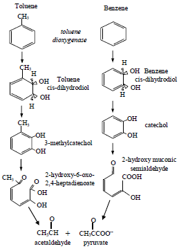
TRIGUEROS, D.E.G.; MODENES, A.N.  y  RAVAGNANI, M.A.S.S.. Biodegradation kinetics of benzene and toluene as single and mixed substrate: estimation of biokinetics parameters by applying particle swarm optimization. Lat. Am. appl. res. [online]. 2010, vol.40, n.3, pp. 219-226. ISSN 0327-0793.

As you can see, toluene can be converted to acetaldahyde and pyruvate (can you then show how both will be used in central metabolism?).

At this stage of your academic career, you should begin getting use to pathway diagrams; not only following them but understanding what is happening.  Microbes, bacteria, archaea, and fungi all have unique and interesting metabolic pathways, and to understand them fully, you must be able to understand their metabolism.


Daily Challenge

Why you studied glycolysis, you studied the most common form, known as the Embden–Meyerhof–Parnas (EMP) pathway.  The Entner-Doudoroff pathway is a variation of the standard glycolytic pathway that can be found in bacteria genera such as Pseudomonas Escherichia, and Enterococcus.  The lactic acid bacteria (a group, or more specifically, a clade of Gram positive bacteria) make use of a pentose-phosphate pathway during catabolism.

Compare and contrast the Entner-Doudoroff and Pentose-Phosphate pathway with the typical EMP pathway you have previously learned.  What are the advantages of these other catabolic pathways, and what are the limitations?  What are important reactions, and why would certain organisms preferentially use these?


Monday, January 27, 2014

BOLO Microbiology Daily Newsletter January 27, 2014 - Metabolism Basics

Site LogoMicrobiology Daily Newsletter

January 27, 2014 - Metabolism Basics


Daily Topic: Metabolism Basics
Metabolism is something all students should be familiar with by this point, especially the catabolic processes of gycolysis and citric acid cycles.  Today, as a preparation for the work this week, we will do some reflection and clarification on catabolic metabolic processes.

1.  What is the purpose of metabolism?  If you say to gain energy, you are only half right.  Catabolism is going to have two outcomes:  energy harvesting and production of precursor metabolites.

The processes of glycolysis and citric acid cycle are going to release energy in the form of reducing potential, and many cells are built to harvest that reducing potential.  But the chemical intermediates of the two processes are also critical for building biomolecules.  We call these two processes "central metabolism" because we can build nearly every biomolecule from the precursor metabolites produced.
Cells have to maintain a balance between energy and precursors.  This is one reason why cells never reach the theoretical maximum yield of ATP production.

2.  What is the energy we harvest from these processes?  If your answer is ATP, then you need to reconsider.  ATP does not equal energy to a cell.  ATP is used to phosphorylate structures.  The -2 charge of phosphate will change the electrochemistry of any molecule it attaches to, and in the case of proteins, will induce a change of shape (conformation).  What then is the energy of cells?  Reducing Potential.  Those electrons moved during redox reactions constitute the real energy harvested during glycolysis and citric acid cycle.  It is the reducing potential we will use in electron transport chains to a proton motive force (an electrochemical gradient of hydrogen ions).

In the eukaryotic mitochondria, this proton motive force will be used to make ATP, but not so in bacteria.  Bacterial proton motive force can be used for active transport and flagellar movement directly.  ATP will still be made, as you need the ability to phosphorylate compounds and proteins, but bacteria will make less ATP than eukaryotic mitochondria.

3.  Why does it take 10 steps to break glucose into pyruvate?  During each step of glycolysis, you are inducing minor molecular changes that induce stress in the molecule.  Through these molecular alterations, the cell carefully extracts energy.  If there was a large change, you risk destabilizing the molecule and releasing energy as heat, which is dangerous to the cell.  The cell needs a series of controlled reactions to capture the most energy.


Words of the Day:
There are four words that you need to know when dealing with metabolism.  The terms deal with how energy and carbon are acquired by a cell.

Energy Acquisition:  These terms deal with how the cell acquires reducing potential.
  •      Phototroph
    • The cell uses photons to raise the energy state of electrons.
    • The cell then transfers these electrons to electron carriers and carbon.
    • In essence, they produce reduced compounds from photons.
  •      Chemotroph
    • The cell uses reduced chemical compounds.
    • The term can be divided into two subcategories:
      • Chemolithotroph - use reduced inorganic compounds.
      • Chemoorganotroph - use reduced organic compounds.
Carbon Acquisition:  This is how the cell acquires carbon for the production of biomolecules.
  • Autotrophic
    • The cell has the ability to take Carbon Dioxide and reduce it to form glucose or another sugar.
    • The process is known as carbon fixation.
    • The cell provides the organic structures needed by the cell.
  • Heterotrophic
    • The cell uses organic structures (or reduced carbon) from external sources (e.g., glucose, amino acids, lipids).
    • These organic structures are converted to other biomolecules as needed.
Each organism is given a name based upon it's principle means of carbon and energy acquisition.  For example:

  • Photoautotroph
    • Photons used to raise energy states of carbon compounds.
    • Cell can fix carbon
  • Photoheterotroph
    • Photons used to raise energy states of carbon compounds.
    • Cells acquire organic structures from external sources.
  • Chemolithoautotroph
    • Reduced inorganic compounds primarily used for reducing potential.
    • The cell can fix carbon.
  • Chemoorganoheterotroph
    • Reduced organic compounds primarily used for reducing potential.
    • The cell acquires organic structures from external sources.


Daily Challenge:  Reflection
What do you remember of metabolism?  Describe what you remember.  Does the information in this newsletter place metabolism into a new perspective for you?  How?  Do you remember the terms listed above?  Describe what you remember.  How do these terms help us to classify cells?  Reflect on this statement:  That the metabolic function of a cell is the most important aspect of a cell.

Thursday, January 23, 2014

BOLO Microbiology Daily Newsletter January 23, 2014 - Bacterial Cell Walls

Site LogoMicrobiology Daily Newsletter

January 23, 2014 - Bacterial Cell Walls


The bacterial cell wall serves the same basic purpose as all cell walls, protection from hypotonic shock.  This protection is achieved by defining the shape and maximum size (volume) of the cell.  The cell membrane can swell to this set limit, but not beyond, thus preventing osmotic lysis.  Due to this hypotonic tolerance, bacterial cells can store higher concentrations of internal solutes than eukaryotic cells, which is advantageous to cells that lack internal compartmentalization.

A defining characteristic of Domain Bacteria is that bacterial cell walls are composed of peptidoglycan.  Glycans are large sugar chains, in this case made up of repeating N-acetylglutamic acid (NAG) and N-acetylmuramic acid (NAM).  Glycan chains are held together by peptide bridges.  Thus, we have peptidoglycan (peptide linked glycans).  In the image below, note how the oligopeptides (that make the peptide bridges) are attached to NAM.  Do you see how this resembles a chain link fence or chicken wire?

Chicken Wire and Chain Link Fence.
Peptidoglycan: http://en.wikipedia.org/wiki/File:Mureine.svg
The following image provides an overview of peptidoglycan synthesis in gram negative cells.  Note that this is a process started within the cytoplasm, but finished outside of the cell membrane.  It is because of this multistep synthesis that peptidoglycan can be targetted with antimicrobial drugs.
Peptidoglycan Synthesis:
Typas, A., Banzhaf, M., Gross, C.A., and Vollmer, W. (2012). From the regulation of peptidoglycan synthesis to bacterial growth and morphology. Nat Rev Micro 10, 123–136.


Since peptidoglycan is defining for bacteria, and not found in mammals, it makes an excellent target for drug therapies aimed at bacterial pathogens.  When you look at medical microbiology and drug development, you will note that a critical stage of developing safe drugs is to target something that is unique to the pathogen.  In this way, you can prevent host toxicity (beyond potential allergic effects).  Targeting a metabolic feature (like glycolysis) that is held in common would hurt the host as well as the pathogen.  [What drugs target peptidoglycan?]

The cell wall structure of bacteria takes on two major forms, referred to as gram + or gram -.  This designation comes from the result of Gram's Staining, a differential staining technique.  Gram + organisms have a large peptidoglycan wall, while gram - cells have a thin peptidoglycan and an outer membrane.  This difference is remarkable, and provides each group of bacteria with its own sets of advantages and limitations.

As a note:  the human immune system has the ability to register peptidoglycan as foreign.   Peptidoglycan is classified as a Pathogen Associated Molecular Pattern (PAMP).  It is picked up by the Toll-Like Receptor (TLR) 2 in human monocytes (innate immunity).  We'll bring this concept up later, but for now, look at the concept of a PAMP...specifically the idea of a molecular pattern. 


Today's Challenge:

Focus today on the gram + cell.  Provide a detailed description of the cell's structure and advantages that can be gained by having a large peptidoglycan cell wall.  Find two examples of gram + organisms, and give some information about these organisms.

Microbiology Daily Newsletter: January 22, 2014 - Bacterial Cell Membranes

Site LogoMicrobiology Daily Newsletter

January 22, 2014 - Archaeal Cell Membranes


Bacteria and Eukarya have a membrane composed of a phospholipid bilayer.  The phospholipid is a common lipid, and naturally gathers so that the hydrophobic tails interact with each other and the polar heads face water.  The lipid bilayer, seen below, creates a perfect semi-permeable structure by having a hydrophobic layer sandwiched between two hydrophilic layers.  NOTE:  The lipid bilayer by its self is semi-permeable; once we add proteins, we describe the membrane as selectively permeable (why?).
Phospholipid Bilyaer: http://en.wikipedia.org/wiki/Image:Lipid_bilayer_section.gif
Like the Bacteria and Eukarya, the Arachaea have a lipid bilayer, but the composition of the lipids is radically different.  What's different?  The phospholipids.

  • Bacteria and Eukarya use an ester linkage between glycerol and fatty acids, while archaea use an ether linkage.
Ester Linked Fatty Acids: http://www.uic.edu/classes/bios/bios100/lecturesf04am/phospholipid.jpg
Differences between Archaeal Phospholipids (Top) and Bacteria/Eukarya Phospholipids (bottom). http://www.ucmp.berkeley.edu/archaea/esterether.jpg
  • Instead of fatty acids, Archaea utilize isoprenoids as the hydrophobic tails.  Unlike straight chain fatty acids, isoprenoid chains have branches, and can include ring structures (cyclopropane and cyclohexane rings are the two most commonly found).  You can see an example of a branched isoprenoid chain in the above diagram.
  • As can be seen above, the glycerol used by Archaea is an enantiomer of the glycerol used by Bacteria and Eukarya.  Archaea utilize L-Glycerol while Bacteria and Eukarya utilize D-Glycerol.  Question: can an enzyme that uses D-glycerol use L-glycerol?
  • In some Archaea, the membrane is composed of a monolayer instead of a bilayer.  In this type of membrane, a long isoprenoid has connects two separate glycerols, each with polar heads.  This type of amphipathic molecule is referred to as a bolaamphiphile.
Archaeal bolaamphiphiles from Ferroplasma acidiphilum.  Pivovarova, T. A.; Kondrat'eva, T. F.; Batrakov, S. G.; Esipov, S. E.; Sheichenko, V. I.; Bykova, S. A.; Lysenko, A. M.; Karavaiko, G. I. (2002). Microbiology 71 (6): 698–706. doi:10.1023/A:1021436107979. ISSN 0026-2617.


Daily Challenge

How would the metabolism of archaeal phospholipids differ from the production of phospholipids in bacteria and eukarya?  What would it take to change from the production of archaeal phospholipids to the production of bacterial/eukarya phospholipids?  How does this change your understanding of the domains of life and the tree of life?

Tuesday, January 21, 2014

Newsletter: Special Edition January 22, 2014 - Prior Knowledge

Site LogoMicrobiology Daily Newsletter

Special Edition

January 21, 2014 - Prior Knowledge


An important activity at the start of each semester is to assess your prior knowledge, and consider your academic strengths and weaknesses. In class today, we reviewed important details regarding cells, and started looking at prokaryotic cells. How comfortable were you with the material? Did you remember the concepts that I brought up?

In order to help you assess your prior knowledge, I ask you to review the checklist below and rate yourself. As a general guideline, at this academic level, you should have a mental construct/picture of the following structures and processes, and have the ability to communicate your understanding. This would be a rating of "Comfortable". If you have an idea about the structure or process, then you would use the rating of "Uncomfortable." If you have no idea about it, rate yourself as "Unaware."
Comfortable Uncomfortable Unaware Topics
                                                Cell Theory
                                                Fluid Mosaic Model of the membrane
                                                Transport Proteins
                                                Receptors
                                                Enzyme Kinetics (Michaelis–Menten kinetics)
                                                Redox Reactions
                                                Electrochemical Gradients (especially the Proton Motive Force)
                                                Replication, Transcription, Translation (central dogma)
                                                how enzymes work
Spend some time to brush up on things you feel weak about.

BOLO Microbiology News Letter: January 21, 2014 - Prokaryotic Cell Membrane

Site LogoMicrobiology Daily Newsletter

January 21, 2014 

The Prokaryotic Cell Membrane


At first glance, the prokaryotic cell shows less complexity than the eukaryotic cell.  The obvious two differences are size and absence of internal membranes.  These two aspects of cells actually go hand in hand.  It deals with the concept of the surface area to volume ratio.  Basically, because prokaryotic cells lack internal membranes, their size is limited.  Eukaryotic cells on the other hand can be larger because of internal compartmentalization. 

But why is surface area so important?  Two reasons should come to mind:
  1. the ability to acquire nutrients and release waste.
  2. the membrane is a key player in many metabolic functions (e.g., electron transport chains).
 In general biology, the cellular membrane (and internal membranes) are focused on because of their importance.  They are the defining structure of the cell.  The cell membrane defines the internal vs. the external environment.  The types of receptors, channels, pores and enzymes a cell puts on their membrane shows the capabilities and metabolic features of a cell.  For instance, if a cell is going to use lactose, it has to have a mechanism to bring lactose across the cell membrane.

Also remember that cells will create concentration gradients across membranes.  You may remember the Sodium/Potassium gradient or the Proton Motive Force.  Both of these are electrochemical gradients, and are critical to the survival of cells. 


Today's Challenge:  Cell membrane structure

You may recall that the cell membrane is described as a fluid mosaic:  Proteins floating in a sea of lipids.  Today, think back on the membrane.  Why is the membrane so critical to the life of the cell?  What reactions have to take place around the membrane?  How important is membrane composition?  What happens when you change fatty acids, or in the case of Archaea, the type of lipids used in the membrane?  What can we learn from the study of membranes?  The above questions do not have to be answered individually.  They are there for you to consider as you build your forum reply.


For more information on cell membranes, go to the Essentials of Cell Biology eBook published by Scitable by Nature.
For more information on archaea, look at the Encyclopedia of Life article on Archaea.

Daily Newsletter November 13, 2013 - Lac Operon part 2

Site LogoDaily Newsletter

November 12, 2013 - Lac Operon


The Lac operon has two levels of regulation.  The first is through a repressor protein that binds to the operator of the operon.  The repressor is constitutively expressed, and binds to the Lac Operon operator.  When lactose is present, it binds to the repressor, thus changing the shape of the repressor protein; the repressor detaches from the operator, and transcription can occur.

There is a second regulation system for the Lac Operon, and it has to deal with the promoter and the CRP binding site.  In the case of the Lac operon, the promoter is "weak".  RNA polymerase does not readily bind to the promoter.  To assist in binding RNA polymerase, there is an activator site (CRP binding site) that can be used to "enhance" the promoter (enhance the binding of RNA polymerase to the promoter).

CRP stands for cAMP Regulator Protein (it is also known as the Catabolite Activator Protein).  CRP has a binding site for cAMP, and the protein is activated (turned on) when cAMP binds.  The CRP-cAMP complex can bind to the CRP binding site, and alter the Lac Operon promoter, enhancing the binding of RNA Polymerase to the promoter.

As can be seen in the image above, CRP-cAMP leads to higher levels of operon transcription.  But why this second regulatory system?

Remember that for this cell, glucose is the preferred carbohydrate and energy source (Escherichia coli is a chemoheterotrophic organism).  The first regulatory system for the operon dealt with the presence/absence of Lactose (you only transcribe the operon when lactose is present).  This second deals with the presence/absence of glucose.  Glucose is the primary carbohydrate source, so as long as glucose is present, there is no need to transcribe pathways for secondary sugars.

In bacteria, the movement of glucose across the membrane (remember hexokinase?) inhibits the production of cAMP.  If glucose transport slows dramatically or stops, Adenylate Cyclase begins making cAMP.  So, while glucose available, there is little to no cAMP in the cell.  When glucose is scarce, we see an increase in cellular levels of cAMP.  For bacteria like E. coli, we can see cAMP as a starvation signal.  The CRP-cAMP complex will bind around the bacterial DNA molecule, and activate numerous pathways for alternative carbohydrate utilization.  As with the Lac Operon, these other pathways will only transcribe when the correct sugar is in the environment.

Daily Challenge 

Explain in your own words the evolutionary advantage of having a two-stage regulatory system, as seen in the Lac Operon. In your discussion, explain why it is important to have secondary messages like cAMP act as signals for large environmental changes, such as starvation states.