Friday, September 28, 2012

Daily Newsletter: September 28, 2012 - Metabolic Evolution & Scientific Articles

Site Logo


Today you are going to look over a scientific article dealing with the evolution of metabolic processes. This is the first scientific article assigned, so I want to spend a little time going over how to read a scientific article.
One of the biggest struggles students have in science is reading scientific articles. These articles are dense, meaning they convey a great deal of information quickly. They are intended for experts, who have an understanding of the background, procedures and protocols. As a result, they are an obstacle when someone new to the field tries to figure out what the authors are saying in the paper.
Most people when they come to a scientific article start with the first line and then plow their way through. Novices become quickly overcome by the language, protocols and jargon, and just get frustrated. Experts rarely read the article from start to finish, but instead skip around. They use the break down of the article to focus their attention on what interests them.
All scientific articles start with an abstract. This is a summation of the paper, but be warned, this summation can be misleading. The purpose of the abstract is to provide a brief rundown of the paper so that people who are looking for an background or protocols can determine if the paper will be of use to them. WARNING: the abstract is for experts, not novices. More than one student has been burned by reading just the abstract and thinking they understood the paper.
While this may be heresy to some, skip the abstract. Don't read the abstract when you'rer assigned a paper. Remember, it is helpful to some one searching through articles. If you are assigned a paper by a teacher, skip the abstract. Instead, go right to the paper's introduction.
Scientific articles are generally broken down into section, with the most common being an introduction, methods, results and conclusion(discussion)*. The introduction holds the background for the paper, and generally includes why the author thinks the study is important. You can usually find the authors hypothesis and assumptions (research logic) here as well.
When you start reading the introduction, do not have a pencil or highlighter in hand. Just do a read through. If you don't understand something, skip it and go on. Just make it through once. On a note pad, write your first impressions. What stood out to you? Start a second read through, but this time have a highlighter, pen or pencil. Mark statements (does not have to be a full sentence) that you think are important. Write down any notes. Ultimately, your looking for a few things in the introduction:
  1. Why does the author think this topic is important?
  2. What has led to this current research?
  3. What is the author's hypothesis?
  4. New terms:
    • Since your new to scientific papers, many of the terms will be new.
    • To start, pick three that seem important to what the author is doing.
    • Look them up and make a note of their definitions somewhere on your copy of the article. (a few words will do)
On your note pad, answer the questions above and make notes.
The METHODsection is one of the hardest to read for a novice, because the whole thing is filled with information on the exact procedures used. Unfortunately, this means you need to have background knowledge on how to do most of these procedures. But this is where we start to learn new techniques. On your first time through, just skim over the method section, but you need to come back to it (Just not immediately)
What to look for in the methods:
This ultimately depends on why you're reading the paper. Are you looking for a method? Are you trying to find an experimental protocol? Or are you trying to figure out how the author got their results? So the questions you ask could change depending upon your goals. The paper you have today has a non-standard arrangement, and the experimental protocol section is minimized (it is actually fleshed out in other sections). Generally though, you want to look to answer the following question:
  1. How did the author set up the experiment?
  • Did they use models systems? (Did they go to a location or did they attempt to replicate the system?)
  • What controld did they use?
  • How many replicates did they have?
  • What experimental methods did they use?
Remember: Do not get bogged down trying to figure out their methods. It is OK if you can not fully answer the questions above. For your notes, pick one procedure that they used and look it up. Write down a one or two sentence description of what the procedure is used for and/or how it is done. It is important to remember that we learn procedures from reading scientific papers.
You should be able to trace the RESULTS back to particular methods used. A good author will provide you a story that leads from methods to results, and finally to a discussion about their conclusions. Results sections normally provide just the FACTS (evidence) that was generated from the methods. As the reader, you are looking to see if the results provide evidence supporting or refuting the authors hypothesis. Your also looking to see what the data says to you. Do the results tell you the same thing they "told" the author? i.e., the results will inform (be the foundation for) the author's conclusions. Based on the same evidence, do you reach the same conclusion? Why or why not? *NOTE: The article today combines results and discussions.
Again, you need to have a good background in the methods to understand and interpret results. So, your goal as a novice is to begin looking at the data to gain an understanding of what the data represents. This is about learning to read graphs and charts. Good authors will lead their reader through the data, but authors have different skills at conveying their data. For the paper today, take one graph, and see if you can figure out all that it is trying to convey. Read the results section, and find where the author discusses the graph. What are they trying to say? *NOTE: For today's paper, explain the results shown in Figure 1.
In the Discussion/Conclusion of the paper, the author attempts to tie together their results and present a logical case supporting their hypothesis. The emphasis here is on LOGICAL. How does the author support the hypothsis? What statements are made to demonstrate how the results support the hypothesis?
For the paper today, find a statement that you think shows where the author demonstrates the data supporting the hypothesis. Also, does the author address future directions for the research? Do they make specific claims?

Reference

Huber, C., Kraus, F., Hanzlik, M., Eisenreich, W. and Wächtershäuser, G. (2012), Elements of Metabolic Evolution. Chem. Eur. J., 18: 2063–2080. doi: 10.1002/chem.201102914
Go to the GSU Library Homepage. Above the search box on the left, you will see a series of tabs. Click on the Journal tab. Type the word chemistry, and click GO. This will bring up a series of Chemistry Journals. You are looking for Chemistry : a European journal. Click on the Find It @ GSU button. You will need to sign-in if you are off campus; follow the sign-in proceedure.
You will then see a series of links that show the access GSU has to different versions. Click on the Full Text Online link. This will take you to the paper. You will have the option to open the paper as a PDF. This is the best way to get a copy to print or save. Open the paper and start to read and take notes.

Daily Challenge

Read the article listed above. In the forum, write about the article. The specific information you need to add includes:
  1. Why does the author think this topic is important?
  2. What has led to this current research?
  3. What is the author's hypothesis?
  4. Three new terms.
  5. How did the author set up the experiment?
  6. New methods/protocolsa
  7. Analysis of Figure 1.
  8. How does the author link data to support hypothesis (conclusion)?
  9. One specific claim or future direction for research.
Link to Forum

Optional Challenge

Review of Lecture on September 28, 2012.
Link to Forum

Thursday, September 27, 2012

Daily Newsletter September 27, 2012 - Enzymes

Daily Newsletter

September 27, 2012 Enzymes


Enzymes are the workhorses of the cell. They catalyze reactions, meaning they decrease reaction activation energy. It is critical to learn how enzymes function. To start with, here are some things you need to remember.
  • First thing to remember, Enzymes are Proteins!
    • So they are constructed on ribosomes.
    • Their structure is determined by electrostatic interactions.
    • Their shape can change when things bind to it.
    • They can be denatured.
  • Second, they act as catalysts.
    • They lower activation energy by bringing molecules together in the correct alignment, and can induce molecular tension to cause the reaction.
    • Though their shape may change during the chemical reaction, the enzyme is left essential unchanged at the end of the reaction.
Enzymes work by binding the substrate of the reaction, and then inducing molecular tension. Remember, when something binds to a protein, the electrostatic interactions around the protein change, resulting in a conformational (shape) change in the protein. It is this shape change that will induce molecular tension. Molecular tension could be due to physical strain, electrostatic interaction, or even the formation of temporary chemical bonds.

Every enzyme has an active site. This is the place where the substrate(s) will bind to the enzyme. The active site must have a shape that loosely fits the substrate, and the electrochemical pattern of the active site must compliment the electrochemical pattern of the substrate. When they bind, you get an Enzyme Substrate complex. Below is a basic cartoon about the process:
This enzyme-substrate complex is critical, and it is likely that you will see the development of an intermediate during the process. As the substrate "binds" to the active site, the overall enzyme will change shape. This conformational change is part of how the enzyme lowers the activation energy of the reaction. In the case of a synthesis, the enzyme will force the two compounds into close association, while in a break-down reaction, the enzyme may appear to bend the molecule at a breaking point. Here is a quick video that shows the brief conformational change that helps to induce the reaction: http://youtu.be/V4OPO6JQLOE


Conformational changes alone are not the only part in inducing a reaction. You will also find that proteins can have prosthetic groups that aid them in their action. For example, you can a Heme group with Iron that can hold Oxygen in red blood cells. Some digestive enzymes use Chromium to help in their action. Many metabolic pathways will contain Electron Carriers needed for redox reactions.

Enzyme Regulation
Cells are filled with enzymes, and they are constantly working to maintain cellular homeostasis. Some of the enzymes though are produced in an inactive form. We need them, but not always. We are also able, though chemical signals, to turn off some enzymes. Last week, we looked at signal transduction, converting a chemical signal into a change in cellular physiology. We saw the action of protein kinases and other primary and secondary signals. One purpose of the protein kinases was to alter the activity of proteins in the cell.

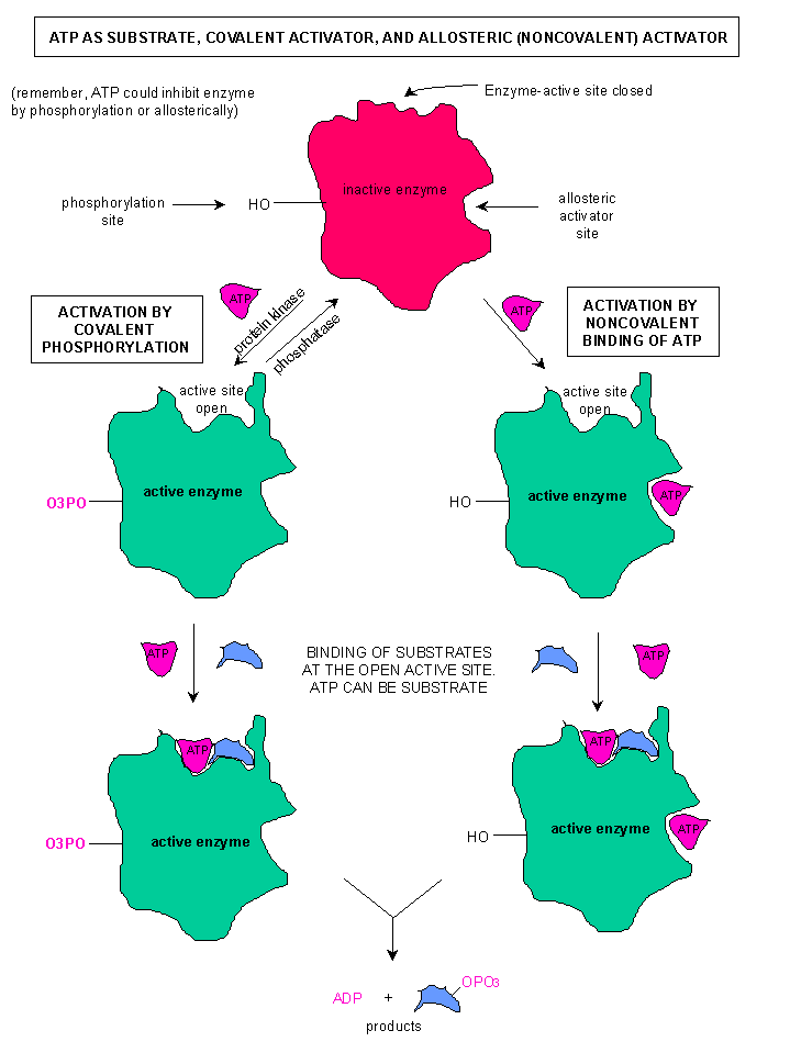
Beyond an active site, many enzymes have an allosteric site (regulatory site). This is located away from the active site. When the regulatory compound "binds" to the allosteric site, the enzyme changes shape. Most critically, the enzyme alters the shape of the active site. An allosteric activator is a signal that opens the active site, while an inhibitor closes the active site. The image to the right shows an enzyme in the active and inactive state. Notice that the active site changes when the allosteric activator is bound to the enzyme. In the case of this enzyme, there are two different ways to regulate: Phosphorylation, which would have occured due to a chemical signal such as cAMP, and the binding of ATP at an allosteric site.

Enzyme RegulationIt is not uncommon to have multiple ways to regulate an enzyme, and some enzymes that can be activated and deactivated as needed. A good example of this is with the enzyme Phosphofructokinase, a critical enzyme in glycolysis. This enzyme catalyzes an energy consumptive reaction that is a "no turning back" point in glycolysis.

Before we talk about phosphofructokinase, we need to talk a little of why we regulate proteins. Two memes I want you to remember: Cells are masters at energy conservation & Cell do not carry out unneccessary reactions. Both concepts are related, and they will help you understand why proteins are regulated. While there are energy consumptive pathways, the main reason we regulate proteins and pathways is due to products. Remember our discussion of equilibrium. We need to use products (products must become the next substrate). If we build up product, the reverse reaction becomes more likely. To ensure that we do not shift equilibrium, we try to avoid product build up. So, we want to stop reactions when we have an adequate to abundant supply of a compound.

The goal of our energy harvesting pathways, of which glycolysis is part, is the production of ATP. So ATP becomes a regulator. Phosphofructokinase has an allosteric site that is specific for ATP. When there is an abundant supply of ATP, some of the ATP binds to phosphofructokinase and changes its shape. Specifically, it closes the active site. PhosphofructokinaseThe presence of AMP in the cell is a singal that we need to unlock our energy harvesting pathways, specifically, unlock phosphofructokinase. In this case, AMP acts as an activator to reverse the inhibitory effect of ATP. To the left is a diagram of phsophofructokinase. The area labeld "Regulatory Site" is the Allosteric site.
In lecture, we will discuss inhibition at the active site.

Additional Resources

Enzymes at Blobs.org - This site has a good review of enzymes, including regulation, inhibition, kinetics and enzyme function at different temperatures. The images presented on the site are extremely helpful.

Daily Challenge

Discuss the concept of enzymes using angiotensin converting enzyme (ACE) as your example. ACE is a medically important enzyme that is involved in an imporant signal system in the body that helps to regulate water balance and the kidneys. There is a group of drugs known as ACE inhibitors that affect the activity of this enzyme. Your goal is to discuss the enzyme, including its regulation and inhibition.
Link to Forum

Wednesday, September 26, 2012

Daily Newsletter: September 26, 2012- Redox and Coupled Reactions

Daily Newsletter

September 26, 2012

Redox and Coupled Reactions


Energy harvesting will be our topic next week, so I wanted to spend a moment and talk about energy in biological systems. Energy is a word that is often thrown around in various disciplines, and most of us carry misconceptions about the world from colloquial (common) use of the word. Scientists define energy as the capacity to do work. This definition helps to simplify a complex issue, but it starts to get confused when we apply it to the complex sets of reactions that we see in living systems.
For example, as we saw yesterday, the work in phosphorylation occurs in creating the covalent bond between a phosphate group and a substrate, not the action of the substrate. With ATP, the emphasis was shifted from seeing ATP as a battery that powered reactions. Instead, your focus was drawn to the Phosphate group, and the electrostatic effect it would have when added to a molecule or protein.

Next week we will discuss Energy Harvesting. Like with ATP, I want you to focus your attention on a specific form of energy, instead of holding a nebulous concept. Today the focus will be on reducing potential. Central metabolism describes the oxidation of glucose, so what are we harvesting? Reducing potential. So what is reducing potential?

A simple definition is reducing potential describes the capacity of a compound to donate electrons. Chemistry has a strict definition involving measurements with electrodes, but for our purpose, the concept of donating electrons is what is important.

Remember the characteristics of life. You must maintain homeostasis, and this means repair. You have to build nucleic acids, lipids, carbohydrates and proteins. These biosynthetic pathways often require you to reduce substrates. To stay alive, you need a constant supply of electrons for reduction; you need reducing potential. If you don't get these high energy electrons for reduction, you die. We will also find that this reducing potential is needed for us to make ATP.

Redox reactions are vital to our survival. Redox reactions are coupled Oxidation and Reduction reactions. One compound is oxidized as the next is reduced.
Remember, the molecules undergoing redox have to be close/touching. But in relative size, a cell is huge compared to a simple molecule. We may harvest electrons (oxidation) in one part of the cell, but use the harvested reducing potential in another part of the cell (reduction). Remember, you don't have free electrons; you can't throw electrons across the cytoplasm. So, how do we couple reactions that may be separated spatially? We use carriers!
Nicotine Adenine Dinucleotide
Electron carriers, like nicotinamide adenine dinucleotide (NAD+), accept electrons at the site of oxidation, and then donate electrons at the site of reduction. NAD+ is readily oxidized and reduced during metabolic reactions, and there is only a negligible loss of energy from the electrons carried (can we ever have NO loss of energy? why or why not?).
NAD+ is also classified as a coenzyme, meaning it must work with an enzyme to accept or donate electrons. NAD+ can not randomly go to a molecule and oxidize or reduce it; its action is regulated by enzymes. NAD+ then must bind to an enzyme that catalyzes an Oxidation, and NADH must bind to an enzyme that catalyzes a Reduction.


Specifically, we couple the reactions. NAD+ has a place to bind into the enzyme, many times next to the substrate. The NAD+ can then capture the eletron pair that is released from the substrate. Additionally, one of the hydrogens will bind to the electron carrier. When we move to the next reaction, NADH will bind with an enzyme, again normally next to the substrate in question. The NADH can then donate the electrons (and hydrogens to the substrate. Enzymes thus help to couple these reactions. NAD+ will not just pick up an electron from any source, and NADH will not just donate electrons to any source. It must be mediated by enzymes.

Daily Challenge

Action of nicotinamide adenine dinucleotide (NAD)
In the citric acid cycle is the following reaction:
In this reaction, malate is oxidized. How do you know? You know because NAD is reduced to NADH. Below is a ribbon model of the protein malate dehydrogenase. Within the protein, you will see two molecules of NAD represented as balls. NAD binds to the enzymes active site first, and then malate binds. Within the active site are both + and - amino acids.

Your task today, using the enzyme malate dehydrogenase, explain how enzymes work and explain how reducing potential is harvested from organic compounds.
Link to Forum

Tuesday, September 25, 2012

Daily Newsletter: September 25, 2012 - Adenosine Triphosphate

Site logoDaily Newsletter

September 25, 2012 Adenosine Triphosphate


Administrative Note

Remember that Milestone Exam 1 ends tonight at 6pm. Make sure you have completed the exam by 6pm.

You have most likely heard ATP referred to as the "energy currency" of the cell. In fact, your textbook uses this analogy: "Just as it is more effective, efficient, and convenient for you to trade money for a lunch than to trade your actual labor, it is useful for cells to have a single currency for transferring energy between different reactions and cell processes."
This is a lovely fiction that does not serve molecular biologists. It is a convenient expression, but it conveys a very serious misconception.

Nucleotide triphosphates (ATP, GTP, CTP, TTP and UTP) have their foundation in the nucleotide structure, with the addition of extra phosphate groups. Adenosine Triphosphate is the most prevalent nucleotide triphosphate, and is found as a cofactor in a number of enzymatic reactions. The picture below show the general structure of ATP.

NOTE: the nucleotide triphosphates use RIBOSE as the sugar. If the nucleotide triphosphate utilized deoxyribose, we add the letter "d" in front of the abbreviation; so dATP represents a deoxyribo-nuclotide. The only time the cell ultilizes dATP, dGTP, dCTP and dTTP is during the process of replication (DNA synthesis).

You have three phosphate groups, each with a negative charge, covalently bonded to each other. The phosphate groups naturally want to repel each other, but they are held together by one of the strongest bond types (covalent bonds). What does this mean? Molecular tension! But it must be noted that ATP is chemically stable. It does not spontaneously loose phosphates (if it did, you would also release heat). It takes enzymatic action to remove the phosphate. When a phosphate is removed from ATP, it is generally attached to another molecular structure (enzymes, sugars, etc...). The exception to this will be in building nucleic acids.

Chemist vs. Biologist

In yesterday's newsletter, a distinction was made between the perspective of chemists and biologists. If you look in most books that deal with biology, you will find the following euqation for ATP:
ATP + H2O → ADP + Pi ΔG˚ = −30.5 kJ/mol (−7.3 kcal/mol)
This is a look at ATP hydrolysis in isolation. Do you remember how ΔG is calculated? Look at the units. kJ (kilo-Joules) or kcal (kilocalories). We are basing this on an isolated reaction and measuring heat. When you look at the ΔG when there are metal ions present, you get a different number. You should also be familiar with ΔG, or Gibbs Free Energy, which is a measure of the amount of work that can be acheived by the energy release. A ΔG˚ = −30.5 kJ/mol is big, and implies that there is a great deal of energy released (the molecule can perform work).

Biologists though recognize that ATP is not in isolation. The intracellular fluid compartment (cytosol) contains ions, and more importantly metal ions, especially Mg2+ (causes major changes in hydrolysis ΔG). Inside of cells, the ΔG˚ of ATP hydrolysis is higher, approximately -50 kJ/mol (-12 kcal/mol). If we released this much energy every time ATP was hydrolyzed, the cell would boil! But, we use this energy for something else: binding the phosphate to another substrate (phosphorylation). When we phosphorylate a compound, we are building a covalent bond between the phosphate and the compound. This requires energy.1 About half the free energy is going to be used to make this covalent bond between the phosphate and the substrate. What happens with the rest? The second law of thermodynamics tells us that some of it is lost (this is an energy transfer after all...we broke one bond to build another), but the bond it self is hold some as well. Remember, chemical bonds are Potential Energy. When phosphate is removed, there will be another change in free energy (every reaction has a ΔG).

So what is the misconception with "energy currency"?
To answer this, we need to ask two other questions: why do we think of ATP as having High Energy Bonds and what is enery to a cell?

Why do we think of ATP in terms of High Energy Bonds? The answer is in the discussion above. The phosphoanhydride bonds (covalent bonds between the phosphate groups) have a high ΔG when they are hydrolyzed. This is why they have been called "high energy bonds". But in biology, the energy is used to immediately allow the phosphate group to bind to a new substrate (Phosphorylation). Put another way, the work that is done is in forming the covalent bond between the phosphate and the new substrate! The ATP is NOT a battery that energizes a curcuit.

What is energy to a cell? Ask yourself, in building bonds in chemistry, what is the energy? If you want to change the energy state in a molecule, what do you do? Isn't it all about the electrons. Ultimately, the energy cells really use will be found with electrons, specifically through redox reactions. We will see that reducing potential is the energy the cells are harvesting and storing. This will be our discussion tomorrow.

Now we come to the big question: What does ATP do?
The concept of ATP as an "energy currency" comes from ATP turning on enzymes or assisting an enzyme during a "power steps" in a metabolic pathways. But ATP does not add energy; it just rearranges charge distribution around a molecule (electrochemistry). Remember, the phosphate group is negatively charged (-2).
When you add a phosphate group to a protein, you change the electrical signature around that portion of the protein (same will be true of other molecules as well). This includes the ability to form new hydrogen bonds, which can alter both secondary and tertiary structures. What will happen to the protein? It will change shape (conformational change). The work that ATP enables (and again this relates back to the free energy) is it's ability to cause conformational changes and induce molecular tension (instabilities).

Protein Phsophorylation
Above  is a general diagrapm showing a phosphorulation event. Notice how the phosphate group has interacted through van der Waals forces to change the tertiary structure of the protein.

The image below is a variant example of how ATP interacts with proteins. In this case, we see the the interaction of Myosin and Actin during muscle contraction. Focus your attention on the myosin molecule in blue. Notice that the "head" of the myosin changes conformation (shape) as ATP binds and is hydrolyzed. This is an important concept. When ATP binds, and hydrolysis occurs, a conformation change occurs. When ADP and Pi leave, the molecule resumes its "resting" conformation. In this case, it is the binding of the entire ATP molecule (with multiple negative charges) and subsequent hydrolysis which causes the conformational change. It still comes down to changing the electrical profile of the molecule (NOTE: there are more conformational changes occuring than are seen in this).  Myosin Cross Bridge (Moving Myosin Heads Bind to Actin)
.

As we will see in upcoming weeks, this change of shape is critical to enzyme function. You have already encountered this once before, with the Sodium/Potassium ATPase.

Daily Challenge

Today, I want you to discuss the function of ATP. Do not describe it as an energy currency, instead describe how the addition of phosphates cause a change in the electrochemistry of a protein, and how that affects the conformation of the protein. Use Myosin in muscle cells as your example. We have not covered Myosin, but it is a very easy model for how ATP acts.
Link to Forum

Reference

1. Berg JM, Tymoczko JL, Stryer L. (2002). Biochemistry. W H Freeman, New York. http://www.ncbi.nlm.nih.gov/books/NBK22399/, accessed on September 25, 2012

Monday, September 24, 2012

Daily Newsletter: September 24, 2012 - Thermodynamics

Site LogoDaily Newsletter

September 24, 2012 Thermodynamics


Modern biology is based on chemistry. What makes up cells? Chemicals. You have water, ions, proteins, carbohydrates, lipids, nucleic acids, and other compounds. Together, all of these chemicals make up what we see as the cell; but remember, the cell is greater than the sum of its chemical parts. It is the interaction of these chemical parts that makes the cell unique.
Biologists look at the chemistry of the cell from a different perspective, or lens, than chemists. In order to understand each individual chemical reaction, chemists will isolate a specific reaction, measure it, manipulate it, and report on it. Our knowledge of biochemistry and molecular pathways are all based on this reductionist perspective (reductionism - simplifying complex systems to component parts for study). Biologists take the information on these parts and attempt to visualize the whole from the individual parts. Due to this, we have to look at how the perspective of biologists differs from that of chemists when we start discussing cellular energetics and chemistry.
Today you are asked to reflect upon the topics of thermodynamics and equilibrium as they pertain to biology. These are topics originally introduced in chemistry, so you may want to go back to your chemistry books to refresh your memory. While biochemists may use these concepts unaltered from their original meaning in chemistry, most biologists look at these two concepts from a slightly different perspective.

The Laws of Theromodynamics: (the two important ones for biology are in bold).
0. If two systems are in thermal equilibrium with a third system, they must be in thermal equilibrium with each other.
(seems obvious to us know; sometimes shows up in biology).
  1. Energy can neither be created nor destroyed, but can be changed from one form to another. (First Law)
    • We are constantly moving energy around in living systems.
    • The most obvious example of this will be the conversion of Light Energy to Chemical Energy in the production of Glucose.
    • Examle: Phototrophs are organisms that can convert light energy to chemical energy.
    • Example: Motile organisms (such as animals) can convert chemical energy into mechanical energy.
    • Example: Luminescent organisms can convert chemical energy into light energy.
    • Example: Heterotrophic organisms can convert one type of chemical energy into a different type of chemical energy. (Remember the First Law includes the conversion of Chemical Energy into a different "form" of Chemical Energy.)
  2. In all energy exchanges, if no energy enters or leaves the system, the potential energy of the state will always be less than that of the initial state. (Second Law)
    • This is big for biology, but not often discussed.
    • You will remember this as the law of Entropy.
    • Every time we experience an energy exchange, some of that energy is used, but the rest is lost due to entropy.
    • Let's say you eat a muffin: Only a fraction of the chemical energy in the muffin will be harvested as useful energy by your cells. The rest is lost to entropy.
    • Heat is a common disorganization of energy, i.e., entropy; but we don't want to over heat cells. (How do we get around this?)
  3. All processes cease as temperature approaches absolute zero. (not generally a concern to biologists).
Later in the week, we will discuss redox reactions, but for now I want to put this concept into your heads: One of the most critical energy exchanges in biology will be the production of reducing potential.

The second law of homeostasis plays a role in homeostasis. With the second law, we know that energetically, we can never break even. Every time that we undergo a chemical reaction, we loose energy. This loss is generally going to be as heat, which is also not good for a cell (too much heat, and the cell boils). So we have be as efficient as possible knowing that we are always loosing energy. How does this play out?
  • Plants take in sunlight, and make glucose.
    • The sunlight is high energy.
    • The glucose is going to have to have less energy because we used photons to excite electrons (an energy change).
  • Animals eat plants.
    • We catabolize the glucose for energy (reducing potential).
    • Do we get 100% of the energy in glucose? NO.
  • Animals build biomass (make proteins, lipids, etc... as needed for life).
    • The energy we got from glucose is further lost when we make new biochemicals.
    • We constantly need energy to keep rebuilding ourselves.
    • We constantly need energy inputs to maintain homeostasis.
So why is the second law so important? To answer this question, we need to look at chemical equilibrium. Chemical EqulibirumYou will recall this concept from chemistry, and it deals with reactions. On the right is an image of an Iron Thiocyanate reaction that was produced by the BBC. It shows the forward and reverse reactions. As you will remember, at a given set of conditions (such as a constant temperature and pressure) you can expect to see an equilibrium reached in which you will have some quantity of substrate and product. While the forward and reverse reactions will continue to occur, there will be a fairly constant quantity of substrate and product.
You may recall that one way to influence the equilibrium is to either add more substrate or remove the product. The removal of product is important for biologists, and sets up a very important condition.
In metabolic pathways, the product of one reaction becomes the substrate for the next reaction. We are constantly taking products to the next reaction. There is a constant flow, at least while an organism is alive, moving product to become the substrate of the next reaction. In biology, it is not about a single reaction, it is always about a series of reactions. This is a critical point! A chemist may look at a single reaction, but a biologist must look at the overall set of reactions if they are to understand the organism.
So, an organism is constantly alterating the equalibirum of a given reaction by taking the product and using it as a substrate in another reaction. We will also see that organisms are constantly acquiring energy and "building block". As such, organisms never acheive the equilibrium a chemist would see in a test tube, and we constantly acquire energy to avoid entropy (consider starvation where you have no new inputs of energy, what happens to the system?).

Daily Challenge

 Today you are tasked with describing in your own words how the laws of thermodynamics and eqiulibrium play out in living systems. Use the above a a jumping off point, but build your own discussion, examples or analogies.

Link to Forum

Friday, September 21, 2012

Site logoDaily Newsletter

September 21, 2012 - Signal Amplification


Resource

Signal Amplification - An animated tutorial produced by McGraw-Hill Publishing. (click on the link)
Secondary Messangers - A section from Cell Molecular Biology by Lodish H, Berk A, Zipursky SL, et al. (2000) from the National Center for Biotechnology Information's Bookshelf. (click on link)

Another important secondary messanger system is the IP3 (Inositol 1,4,5-triphosphate) pathway. A primary purpose of this pathway is to release Ca2+ that is stored in the Endoplasmic Reticulum. Ca2+ becomes an activator of various proteins. An interesting aspect of this system is that IP3 is derived from phosphatidylinositol 4,5-bisphosphate (PIP2), a phospholipid found in cell membranes. PLC cleavage of PIP2 to IP3 and DAG initiates intracellular calcium release and PKC activation. The system then uses a membrane component as the signal agent. The system also requires a second lipid component of the membrane known as diacylglycerol (DAG). To the right is a general schematic of the system from Wikipedia Commons.
 
Phospholipase C is the enzyme that converts PIP2 into IP3. It is also generally the signal receptor. IP3 is water soluble, and will bind to the IP3 receptor on the Endoplasmic Reticulum, opening CA2+ channels. Calcium ions are then able to activate Protein Kinase C with the help of DAG. At this point, we have phosphorylation of substrates (mainly other proteins).
The signal increases the presence of IP3 in the cell, which changes calcium ion concentrations in the cells. An increased [CA2+] provides a greater probability that Protein Kinase C will be activated, resulting in a physiological change to the cell.

Daily Challenge

Explain the concept of signal amplification either using cAMP or IP3 as your example system.
Link to Forum
Link to Optional Challenge: September 20, 2012 - Lecture Review Forum

Thursday, September 20, 2012

Daily Newsletter: September 20, 2012 - Direct vs. Indirect Signals

sDaily Newsletter

September 20, 2012 Direct vs. Indirect Signals


Signal transduction involves a signal contacting an appropriate receptor which ellicits a cellular effect. The transduction is thus the changing of signal into an effect. Transduction can be either direct or indirect.
A direct transduction event implies that the receptor ellicits an immediate effect on the cell. A good example of this is the Androgen (Testosterone) Receptor. The image below is a good image of the basic signal transduction pathway. Testosterone is lipid soluble, so can easily move through the membrane. Once inside of the cell, it is changed to dihydroxytestosterone (DHT). This binds to the receptor, and then the receptor-ligand complex is moved into the nucleus. Inside of the nucleus it directly affects the transcription of genes. In this case, the receptor-ligand complex has a direct effect on the cell.androgen receptor
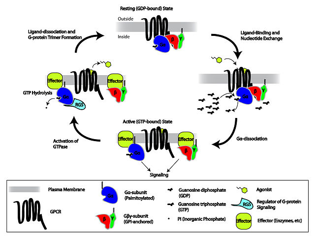
Indirect signal transduction will involve the creation of a secondary messanger. The receptor-ligand complex will create a condition where an intracellular chemical signal is created. One primary signal binding to the receptor can then be amplified within the cell by creating this secondary messanger. Below is a general diagram showing the use of a G-Protein interface. The Ligand-Receptor complex directly activates the G-Protein. The activated complex then activates a specific effector in the cell membrane.
Cartoon depicting the Heterotrimeric G-protein activation/deactivation cycle in the context of GPCR signaling
In the above image, there are four views of the Receptor-G-Protein complex. In the top image of the cycle, the Resting State, there is no ligand bound to the receptor. When the ligand binds (the next image in the cycle), GTP binds to the Gα subunit. This causes the Gα subunit to detach from the rest of the G-protein (Gβγ Subunit). The subunits then bind to Effectors.
A very prevelant effector enzyme is AdenylatAdenylate Cyclase actione Cyclase. This enzyme takes ATP (Adenosine Triphosphate) and removes two phosphates. The remaining phosphate binds back to the ribose sugar at the 3' carbon, creating a ring structure (cycle) between carbon 5' and 3'. This is known as cyclic AMP (cAMP). cAMP is a very common secondary messanger. It can be produced in bulk and can bind to a variety of enzymes. Many kinases (enzymes that add phosphates to other proteins, aka phosphorylation) are activated/deactivated by cAMP. So eukaryotic cells use this system to amplify the original signal to produce a large number of cAMP in order to affect protein activiation.
Why would phosphorylation affect protein activiation? Phosphates are large with a -2 charge. Binding a phosphate to a protein will force the protein to change shape (configurational change).
Below is an example of indirect signal transduction via a G-protein:
Epinephrine-stimulated cAMP synthesis

Daily Challenge

Using Testosterone and Epinephrine as examples, explain the difference between direct and indirect signal transduction
Link to Forum

Reference

The image of Epinephrine stimulated cAMP synthesis comes from Encyclopedia Britanica.
Encyclopedia Britanica. (2008). Epinephrine-stimulated cAMP synthesis. Encyclopedia Britanica, http://www.britannica.com/EBchecked/topic/1522055/G-protein-coupled-receptor-GPCR?overlay=true&assemblyId=124084, accessed September 12, 2012.
Other images are from Wikipedia Commons with the full reference embedded as a pop-out, or have the reference shown in the image.

Wednesday, September 19, 2012

Daily Newsletter: September 19, 2012 - Ligand & Receptor

Site LogoDaily Newsletter

September 19, 2012 Ligand & Receptor


Ligands are chemical signals, and receptors are proteins. The receptor is folded so that it forms a binding space (active or binding site*) where the ligand can dock and bind. A common thought is that every receptor has a specific ligand, and that nothing else binds to the receptor. A lovely fiction.
To the right is an image of the μ-Opioid receptor (μ is the Greek Letter Mu). μ-Opioid ReceptorThis receptor is found in human neural tissue, and provides analgesic effects and feelings of euphoria. It can also cause respiratory depression and reduced GI motility. The main ligands for this receptor are enkephalins and β-endorphin. Yes, this receptor set has two potential ligands. It also has a list of agonistic and antagonistic compounds that can bind to the receptor.
When we speak of receptors and ligands, we talk about the affinity of the receptor for a particular ligand. Remember, we have folded the protein to create a 3-D shape. In the case of the μ-Opioid receptor, the folded protein has a notch where the ligand can slip in. This notch will have chemical properties complementary to the ligand. A high affinity would imply that the physical shape and chemical properties of the notch are a good match to a given ligand. In this case, beta-endorphin is a strong chemical and physical match to the binding site (notch) of the μ-Opioid receptor. Dynorphins, which are another type of neurotransmitter (ligand) in the brain, have a low affinity for the μ-Opioid receptor's binding site, meaning the shape and chemical properties are not a good match.
This sets up another aspect of receptors: agonists and antagonists. These terms represent chemical mimics of the natural ligand(s) of a given receptor. Agonists are chemical mimics that bind to a receptor and trigger a cellular response; in other words they work like the ligand. Antagonists on the other hand are chemical mimics that bind to a receptor and block a cellular response. Active and inactive μ-opioid receptors.In fact, antagonists can stay bound and prevent activation of the receptor. A well known agonist for μ-Opioid receptor is morphine. Morphine can bind to the μ-Opioid receptor and active the cellular response that leads to analgesic effects, feelings of euphoria and respiratory depression. To the left is an image depicting agonistic and antagonistic bindings possible with the μ-Opioid receptor. NOTE: The agonist relationship implies an activation of the cellular response, while the antagonistic relationship implies an inactivation of the cellular response.
Morphine is a powerful analgesic with many well known side effects. One of the most dangerous of these side effects is physical addiction and an increased tolerance for the drug (you need more and more to get the same effect over time). This provides a good example of another principle of receptors: Regulation.
Down Regulation: When a cell receives too much signal, it will begin to down regulate the receptors. This means that the cell stops producing the quantity of the specific receptor it usually makes. For membrane bound receptors, over time, as the membrane is repaired and refurbished (a constant dynamic process), the number of expressed receptors drops. The result is that the cell is less sensitive to the signal. Why does a cell do this? Think of it as a person being exposed to loud noises. If it happens once, for a short period of time, the body can compensate. What if the person is continuously exposed to the loud noise? Eventually they become less sensitive to sound, i.e., they become functionally deaf. The same thing is happening to a cell that is overexposed to a ligand. To protect themselves, they produce less and less of the given receptor. In some cases this becomes an irreversible loss of the receptor from the cell. In the case of the μ-Opioid receptor, your body produces only small temporary doses of β-endorphin. With morphine, you have a large dose, and it is usually for a long duration. The longer the duration (over a week), the more likely you will have desensitization (down regulation) of the receptors.
Up Regulation: The reverse of down regulation is up regulation. If a cell is not getting enough signal, it will start building more receptors. In this case, there is a deficency in the amount of the ligand preset. The cell is compensating by building more receptors.
Both Down Regulation and Up Regulation are examples of Negative Feedback.
*NOTE: Receptors and Enzymes are both proteins. They both have a site where a ligand (receptor) or substrate (enzyme) can bind. As with receptors, we will see that enzymes have an affinity for their substrate, and like receptors, other chemicals can bind into the enzyme. In addition, like ligand-receptor, when a substrate binds into an enzyme, the enzyme will change shape.

Administrative Note

Regarding the Milestone Paper and References, remember that you are to use scholarly sources for your references. Your Textbook is considered a scholarly source for this paper, as are the Daily Newsletters. Your own forum posts are not considered scholarly resources. Use your forum posts to help build your paper's structure, and any references used in making them. DO NOT cite your own forum posts.
Remember that citations are to use the American Medical Association format. If you are familiar with APA, that is an acceptable alternative.

Daily Challenge

Modern drugs tend to act as either agonists or antagonists to specific receptors in the body. This includes both medicinal and illegal drugs. Pick one of the following drugs and discuss the effect it has on its target receptor. Include the effect on the human body. (NOTE: You're not expected to write a massive paper on this; just show an understanding of the concepts presented here and an understanding of the drug you chose to write about).
  • Epinephrine
  • Metoprolol
  • Phentolamine
  • Guanfacine
  • Aripiprazole
  • Quetiapine XR
  • Zolpidem
  • Buspirone
LINK TO FORUM

Daily Newsletter: September 18, 2012 - Basics of Cell Communication

Site logoDaily Newsletter

September 18, 2012 Basics of Cell Communication


Cells must be able to sense their environment, and respond to environmental stimuli. Cells must have some mechanism to receive environmental signals, and process those signals into a response. All cells, prokaryotes and eukaryotes have this ability. Beyond just responding to the environment, we now know that cells, even bacteria and archaea, possess the ability to signal each other. In multicellular organisms, we will talk about the coordination of metabolism and growth using chemical signals. In humans, from the embryo stage til death, are cells are constantly talking to each other.
Signal reception occurs in all known organisms (from picking up environmental to cellular signals). For instance, in bacteria, we know of a signal phenomena known as Quorum Sensing. With this signal system, bacteria release chemicals as they grow which other individuals of the same species (and some times other species) pick up; when the concentration of the chemical reaches a critical point, cellular changes can be observed in the community. Basically, as the population increases, cells begin to change.
But how do cells pick up signals? Signal recognition begins with protein receptors. For a cell to pick up, or register, a signal, it must build a receptor for that signal. (IMPORTANT NOTE: a cell that lacks a receptor for signal X can not register signal X; they are deaf to the signal). This sets up another question: what is a signal?
Most of the signals we will talk about are chemical signals (aka, Ligands), meaning we have a chemical compound that will "fit" a receptor, activating it. There are other signals though: light can be a signal (photoreception), temperature (thermoreception), and even mechanical such as touch (mechanoreception). As mentioned, out discussions for this week will focus on chemical signal pathways.
By far, the most common type of signal system will involve chemicals. Hormones are chemical signals, neurotransmitters are chemical signals, even carbon dioxide is used as chemical signal in the human body. Because there are so many different chemical signals, we have a generic word for any compound that could bind and activate a receptor protein: Ligand. As a general word, ligand is used when we discuss the basic concepts of signal systems. (NOTE: In biology we have a number of GENERIC words that are used in discussing basic pathways or models. Ligand is one of those terms).
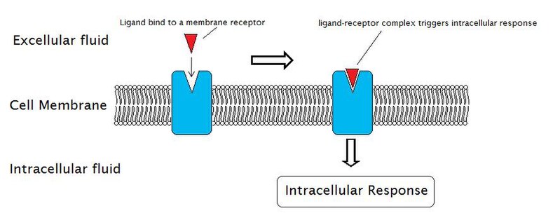
At their most basic, a chemical signal system (or pathway) will be comprised of a Ligand and a Receptor. When a ligand binds to a receptor (ligand-receptor complex), the receptor changes shape (conformation), which elicits a physiological response in the cell. Remember: The receptor is a protein, and when proteins change shape, they have an effect on the cell. So the basic signal system will be: Ligand binds to receptor, receptor changes shape, cellular response occurs. This is known as Signal Transduction.External reactions and internal reactions for signal transduction

Words of the Day: Paracrine & Autocrine

Prepare definitions for these two words and put them in your notes.

Daily Challenge

Below is a diagram of the Insulin Receptor and Signal Transduction. Review the image and information in your text, then write a forum post describing the nature of the Ligand and Receptor, and then the effects on the cell.
LINK TO FORUM
Effect of insulin on glucose uptake and metabolism.

Special Edition: September 18, 2012 - Week 5 Outcomes

Special Edition Newsletter

September 18, 2012 Week 5 Outcomes


Administrative Notes

Apologies for the newsletters starting late this week. There will be four newsletters this week, Tuesday to Friday, with a special edition after the Thursday lecture.
You wil note two new labels in the General screen/tab: Active Quizzes and Current Milestones. All active quizzes and information regarding current milestone assignments will be found in this area.

Learning Objectives: Cell Communication

  • Environment to Cell Communication.
  • Generalized Signal Transduction Pathway.
  • Signal Receptors
    • Ligand-Receptor Complexes.
    • Types of Receptors.
    • Importance of Receptor Location.
  • Importance of Protein Kinases.
  • Importance of Secondary Messengers.
  • Types of Secondary Messengers.
  • Cellular response to primary or secondary messengers.
  • Importance of Gap Junctions.
  • Importance of Plasmodesmata.
Remember that all of these topics may appear on your quizzes or exams.

Friday, September 14, 2012

Daily Newsletter: September 14, 2012 - Evolution Friday

Site logoDaily Newsletter

September 14, 2012 Evolution Friday


The evolution of the cell, especially the development from prokaryote to eukaryote, is an important topic as it demonstrates core differences between living organisms. Archaea, Bacteria, and Eukarya are dramatically different from each other, but even within these domains, there are vast differences in cell types. When studying the development of cellular differences and structures, and thus studying the evolution of cells, biologists often look toward existent organisms that do not fit the typical cellular model. Anomalous structures give us insight into possible evolutionary steps.Symbiodium symbiosis with Jellyfish
For example, the genus Symbiodinium is an algae of the phyllum Dinoflagellata. These algae are known to live inside the cells of cnidarians (corals and jellyfish). Within the cell, they algae will deliver photosynthetic products to the cnidarians. Sound familar? How does this cell "know" to become symbiotic? Are there signals between the two cells? Does this mimic the entry of a Cyanobacteria into a cell where it became the chloroplast?
Then there is the bacterial phyllum Planctomycetes which researchers suspect is the "missing-link" between prokaryotic and eukaryotic cells, i.e., the development of a nucleus. Beyond membrane alterations, this is a bacteria that reproduces by budding (the daughter cell has a reduced cytoplasm). Other bacteria reproduce by binary fission, which results in two daughter cells with equal cytoplasm.Bdellovibrio Life Cycle
The bacterium Bdellovibrio is of interest in that it is a predator of other bacteria. This organism hunts other bacteria, enters their cell wall, then devours them. This is a very unusual characteristic. Not only in the recognition of prey, but in the entire life cycle. This is a complexity that we do not see in other bacterial groups.


Daily Challenge

Review the following diagram showing proposed steps in the evolution of the eukaryotic cell.
Evolution of Eukaryotic Cell
In your own words, discuss the origin of the eukaryotic cell. Using the examples above, and any others you find, how can we provide evidence of these evolutionary steps or stages. Some claim that the Archaea are the ancestors of the Eukarya, but if so, why are there so many differences, espeically when it comes to phospholipids? How easy would it be to change the phospholipids a cell uses?

Link to Forum

Thursday, September 13, 2012

Daily Newsletter: September 13, 2012 - The Mitochondria

Site logoDaily Newsletter

September 13, 2012 The Mitochondria


The mitochondria is known as the power house of the cell, for this is where eukaryotic cells experience oxidative phosphorylation and ATP production. We will come back to ATP in a latter newletter, but you should note that phosphorylation of proteins is a powerful activator of enzymes (allowing them to work). Everything from pump systems, cellular movement, and even muscle contraction relies on ATP.
Like the structure of the nuclear envelope, the mitochondria is a double membrane bound structure, but the origin of the nuclear envelope and mitochondria are very different. It is hypothesized that the nuclear envelope formed from the infolding (invagination) of the cell membrane. The mitochondria, in contrast, is two separate and distinct membranes.
The Endosymbiotic Theory is used to explain the development of the mitochondria. (Question: why should we consider this a theory?) Before we get to the endosymbiotic theory, we need to first look at the structure of the mitochondria:
We have an outer membrane and an inner membrane. Between the two membranes is the Intermembranous Space. The inner membrane is highly folded into Cristae (Question: why would you fold a membrane?). The inner compartment, bounded by the inner membrane, is known as the mitochondrial matrix.
With the structure in mind, how does this differ from the nuclear envelope?
  • The outer membrane displays eukaryotic proteins.
  • The inner membrane displays prokaryotic proteins.
  • The intermembranous space stores hydrogen ions, so is acidic.
  • The matrix contains a circular bacterial DNA molecule and 70s (prokaryotic) ribosomes.
  • The mitochondria is self-replicating (the DNA can make copies).
NOTE: Remember that phospholipid bilayer membranes allow you to create seperate fluid compartments. Here we have three fluid compartments: Intracellular, Intermembranous, matrix. Are these three fluid compartments have different chemical or electrochemical concentrations?
NOTE: The inner membrane seperates the intermembranous space (acid) from the matrix. We find that there is a H+ gradient across this membrane. Pumps along this membrane maintain the gradient by pumping H+ ions from the matrix into the intermembranous space. Pores along the inner membrane allow H+ ions to move back into the matrix. REMEMBER: when ions move down their electrochemical gradient across a membrane, work is done. There is a proton motive force across the inner membrane (this is the name we give to this type of H+ electrochemical gradient. 

QUESTION: Why do we not consider the outer membrane to have an electrochemical gradient?
 
The endosymbiotic theory describes the mitochondria as a bacterial symbiont that was engulfed by a "proto-eukaryotic" cell. A relationship formed between the two cells, with the mitochondria taking over ATP production, and the "proto-eukaryotic" cell loosing the ability.

Genenomic analysis of the mitochondria shows that it comes from the bacterial Order Rickettsiales, which means that it is related to the intracellular parasite Rickettsia rickettsii (Rocky Mountain Spotted Fever). Mitochondrial genes are inherited matrilineally, and are the basis of human population genetics studies of the mitochondrial genome.

Like the Mitochondria, the cholorplast is an endosymbiont that exists in eukaryotic photosynthetic cells (such as plant cells). Photosynthetic cells will have both mitochondria (extract energy) and chloroplasts (build reduced organic compounds). The function of the chloroplast is to use light energy to reduce carbon (carbon fixation) in order to produce reduced carbon compounds, such as glucose. The chloroplast, like the mitochondria, has two membranes, one eukaryotic in structure and ond prokaryotic. The chloroplast is also self-replicating, and has baterial ribosomes and genophore (DNA).


Daily Challenge

Write about the mitochondria, the endosymbiotic hypothesis and human mitochondrial genetics. Explore these topics, and feel free to go deeper on any feature of the mitochondria that interests you. One question I want you to focus on is why is the mitochondrial genome reduced (smaller) than other members of the Rickettsiales? In your discussion, you must answer the question: Why do we not consider the outer membrane to have an electrochemical gradient?
Link to Forum

Wednesday, September 12, 2012

Daily Newsletter: September 12, 2012 - Cytoskeleton

site logoDaily Newsletter

September 12, 2012 Cytoskeleton


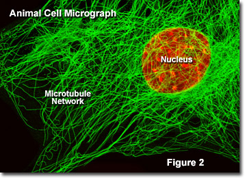
The cell is not just a fluid filled sack, but an organized structure complete with internal supports. Consider it a high rise building that is just squishy. Creating the structure are a protein fibers (structural proteins). Structural proteins typically are in quaternary structures, meaning that individual proteins combine to create the overall structure. The cytoskeleton is composed of Microtubules, Microfilaments, and a group of proteins referred to as the Intermediate Filaments.
The microtubules are one of the most well studied cytoskeletal elements. They are composed of the protein tublin, and are responsible for the flagella, mitotic spindles, internal structure, and movement of materials and vesicles within the cell (they act as roads on which vesicles are carried).
Microfilamentsare composed of actin. They help to form structure, and are used in movement, such as muscle contraction, the formation of pseudopods and bulk transport.


The intermediate filaments are a family of structural proteins that are smaller than microtubules, but larger than microfilaments. There are a number of different types, and some are specific for a given cell type. Wikiepdia provides a good list of the different types of intermediate filaments in the article intermediate filaments.
Spectrin is a structural protein that was originally classified as an intermediate filament. In recent years, the importance of this protein to the cell membrane has become more noted, so it deserves it's own place. The structure of spectrin has it reclassifed as related to actin (microfilaments).

The cytoskeleton plays a major role with movement within the cell, especially when it comes to forming organelles and moving vesicles. We see the cytoskeleton helping to form the nuclear envelop (the nulcear lamella), and are important in the disappearance of the nucleus during cell division. They are also responsible for the shape of the endoplasmic reticulum, formation of vesicles and the Golgi body, as well as the incorporation of vesicles into the cell membrane.

The endomembranous system, which includes the endoplasmic reticulum and Golgi body (apparatus). This system is critical for the proper formation of proteins that will be associated with the membrane, other organells, or secreted from the cell. The rough endoplasmic reticulum (which get's its name due to the presence of ribosomes on the surface of the ER) is the location of protein synthesis for proteins with the above listed fate. The smooth endoplasmic reticulum is a place of protein processing and lipid (especially phospholipid) synthesis. It is in the smooth endoplasmic reticulum that we see the production of new phospholipid bilayer components (i.e., phospholipids).


Endomembranous system
Vesicles from the endoplasmic reticulum will go to join/make the Golgi body, where proteins are further processed, packaged and stored. Vesicles from the Golgi body will then be taken to a destination, either another organelle, an internal vesicle/microbody such as the lysosome, or will be incorporated into the cell membrane. Membrane bound vesicles add new phospholipids (bilayers) with integrated proteins into the membrane. We are constantly adding new material to the cell membrane in this way. REMEMBER: the membrane is dynamic, meaning, we are always adding and removing component pieces. Also remember that it is the cytoskeleton that makes the formation and movement of these structure possible.


Daily Challenge

Today, you are to describe in brief the function of the cytoskeletal elements listed above. You do not need to go in depth about the structure, at present the function is more important. Pick one of the above cytoskeletal elements to be your focus today. Write more about that element, including it's structure. Do not neglect the other components.
Link to Forum

Daily Newsletter: September 11, 2012 - The Nucleus

Daily Challenge

September 11, 2012 - The Nucleus


Admistrative: Milestone Paper

Your first Milestone Paper is due to be loaded next week. Remember that you can use any information written in your daily challenge to help build this paper.

The nucleus defines the eukaryotic cell. Why would we say that? Remember yesterdays discussion about prokaryotes and eukaryotes? The principle differences is the presence or absence of a nucleus. Eukaryotes have a nucleus, while the prokarotes don't. Presence of a nucleus also implies the ability to create internal membranes.
The nucleus is a central structure present in eukaryotic cells, and is the site where we find the cell's DNA. It is a highly regulated structure, and one function is to ensure the protection and stability of the cell's genetic information. We also consider the nucleus an organelle.

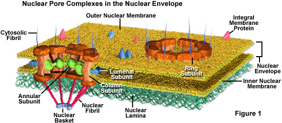
The nucleus is a double membrane bound structure, which means that there are two lipid bilayers that make up the nuclear envelope.The outer layer of the nuclear envelope gives rise to the endomembranous system, which includes the Endoplasmic Reticulum and the Golgi Apparatus.

To gain access to the inside of the nucleus, you must first move through nuclear pores. Remember that phospholipid bilayers are selectively permeable. The nuclear pores are large. The purpose here is not to control ion movment, but macromolecule movement. For example, messager RNA (mRNA) will need to leave the nucleus so that proteins can be made in the cytoplasm. mRNA is a large linear macromolecule, so to move it from the nuclear compartment to the cytoplasm, you have to pass the mRNA through nuclear pores. These pores are regulated so that only specifically tagged macromolecules can move though. So the goal is not to protect the DNA from small molecules, but provide protection from macromolecules. Click on the link for more information on the nuclear pore complex.
Inside of the nucleus, you will find a region known as the nucleolus. In micrographs (pictures generated from microscopes), you will see the nucleolus staining differently than the rest of the nucleus. It appears denser. This is a region of active transcription (making RNA). Ribosomal RNA, Transfer RNA, and Small Nuclear RNA is continually being transcribed (synthesized) in this area, hence the reason for the difference in appearance.
The job of the nucleus is to protect the cells DNA. We will discuss DNA in more detail later, but for now know that it holds the code (in the form of nucleotides) for how to create the different forms of RNA, and thus how to create proteins. Changes in DNA will change the resulting protein's primary structure (sequence of amino acids). As we learned earlier, changes to the primary structure will affect the secondary and tertiary structures, and thus, the function of the protein. DNA needs to be protected from oxidizing agents, digestive agents, and any other harmful macromolecule.
NOTE: prokaryotes do not have a nucleus! The DNA of a prokaryote is found in the cytoplasm of the cell. As DNA has a tendency to stain differently than the rest of the cytoplasm, we can generally visualize the region of the cell where the DNA is found. In prokaryotes, this region is referred to as a nucleoid.
prokaryotic cell

Daily Challenge

In your own words, describe the structure and function of the nucleus, including the action of the nuclear pores.
Link to Formum

Daily Newsletter: September 10, 2012 - Cell Theory and Domains

Site logoDaily Newsletter

September 10, 2012 Cell Theory and Domains


The fundamental unit of life is the cell. This is one of characteristics of life, and is a foundational principle of biology. Based on the discussions from last week, you see that for life to exist, we must first create a barrier that seperates and inside fluid compartment from the outside (external) fluid compartment. This allows us to create electrochemical gradients that will be necessary for life functions. When that membrane seperating inside from outside is disrupted and broken, life functions cease. While the cell membrane may be the defining structure of the cell, the cell is more than just the cell membrane.
This week, we turn our attention to cellular structure, and begin to look at the life functions of a cell. The first step is to recognize that all life is made up of cells, and that all living cells come from pre-existing cells. The following phrase, highlighted in yellow, is the core of the modern cell theory. This theory, which we credit to the work of Matthais Schleiden (1838) and Theodor Schwann (1839), was based upon growing microscopic work in the early 1800's. Work, even into the modern day, has provided robust evidence to support this theory, and has helped in some refinements.
What is interesting about the cell theory are the implications, e.g., a complex organism, such as man, is made up of millions of individual, and seemingly independent, cells. The overall organism, is an expression of the total activity of each of these cells. This has led to people studying communication between cells, for how do you coordinate the action of millions of individual cells? Another study is how the individual metabolic operations of cells can sum to the overall metabolic operation of an organism. As you can see, the idea of the cell being the fundamental unit of life has rather important implications for biology.
When we look at life, we begin to notice that there are different types of cells. The most basic difference between cells is the presence or absence of a nucleus. Prokaryotic cells lack a nucleus, while eukaryotic cells contain a nucleus. The nucleus, which will will cover in more depth later this week, is an internal comparment that contains the cells DNA. An internal compartment means that there is a membrane barrier that seperates the contents of the compartment from the rest of the cell. Like the cell membrane, these internal compartments are surrounded by protein containing phospholipid bilayers (fluid mosaics). As the cell membrane seperates the extracellular fluid from the intracellular, so to do these internal membranes create new fluid compartments. Just like the cell membrane, these internal membranes are selectively permeable, and allow for internal spaces with different chemical concentrations.
The presence or absence of a nucleus is a major taxonomic feature for organisms, and the ability to form and maintain internal compartments has many implications for the organism (some of which we will discuss this week). One of the major differences is in size. Remember all the proteins found on the cell membrane? An important thing to remember is that there are a termendous number of metabolic reactions that occur at the membrane, as well as the import and export of ions. We have to have enough membrane to satisfy our metabolic needs. The larger the volume of the cell, the smaller the surface area, so cells have to remain small. That is, unless you have the ability to make internal membranes. Internal membranes increase the available surface area...so eukaryotic cells, which are able to make internal membranes, can be larger than prokaryotic cells.
Living organisms are divided into three domains: Eukarya, Archaea and Bacteria. Eukarya contains all cells that are eukaryotic (nucleus containing). The Archaea and Bacteria are prokaryotic. The division of organisms into three domains was based on genetic and biochemical analysis conducted over the previous 30 years. Each domain represents a unique cellular structure.

Daily Challenge

Members of all three domains are based on the fundamental unit of the cell. For today's challenge, you are to discuss the differences that led to the three domains being declared unique from one another.
Link to Forum

Reflection

Cells are the fundamental unit of life, but over the past four years, virologists have been saying that the definition of life should be expanded to include acellular structures such as viruses. Currently, viruses and other acellular structures are considered to be biological agents; i.e., structures that can affect the genetics and metabolism of a cell, but are not technically living. What would be the logic behind expanding the definition of life to include viruses? What problems would it create?