Wednesday, October 31, 2012

Daily Newsletter: October 31, 2012 - Codons, Anticodons & Amino Acids

Site LogoDaily Newsletter

October 31, 2012

Codons, Anticodons & Amino Acids


Translation is the process of "reading" mRNA, and using the code to construct a protein. But what is the code? The nucleotide language of mRNA can be divided into codons. Three sequential nucleotides that represent a genetic (nucleotide) word. So, how do you read this code or nucleotide language?
In the image to the right, you hav
e have sequential nucleotides divided up into codons. Notice that AUG is listed as Codon 1. This is important! AUG is the Universal Start Codon. Nearly every organism (and every gene) that has been studied uses the three ribonucleotide sequence AUG to indicate the "START" of protein synthesis (Start Point of Translation).

As we will see tomorrow, it takes more than a start codon to initiate transcription, but for now just remember that this is the codon that indicates the START point of the instructions on how to make a protein.
The start codon established the Reading Frame for translation. From the start codon, every three sequential nucleotides will be viewed as a codon. This is critical! Mutations can affect reading frames. For example, if a nucleotide is inserted between codon 2 and 3 (G G), would you have the same reading frame down stream? What if you deleted the first nucleotide of codon 4? What is the effect of changing the reading frame? What would happen to the resulting protein?

Insertions and deletions can change reading frames, but point mutations can also occur. In this case, one nucleotide is change to a different nucleotide. What would happen if the final nucleotide of condon 3 were changed to a C? To an A? How about the second nucleotide in codon 4? Change the U to an A, what happens?

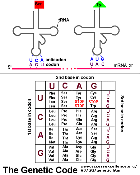
Each codon is a "genetic word," and refers to a specific amino acid (thus changes to these words can result in changes to final proteins). The tRNA is the agent of translation. On one end of the tRNA, you will find an anti-codon. Anti-codons are complimentary to codons. Example: Codon 1 reads AUG. The corresponding tRNA would have an anticodon reading UAC. (Question: Would these be antiparallel?). Codon 2 reads ACG, so the anticodon would read UGC. Oppisite the anticodon, you will find a binding site for a specific amino acid.

An amino acid can be attached to the free 3' end of the tRNA. There is a class of enzymes capable of attaching an amino acid to a tRNA: Aminoacyl tRNA Synthetase. Below is a very basic cartoon of how an amino acid is added to a tRNA.
Note that an ATP is needed to complete the binding. There is an Aminoacyl tRNA Synthetase for each tRNA-Amino Acid combination.
Below is a diagram showing the pairing of codon to anticodon. The diagram also contains a version of the Genetic Code table, showing the relationship between codon and amino acid.
Note that three codons are referred to as STOP codons: UAA, UAG, and UGA. These are used to terminate translation; they indicate the end of the gene's coding region. What would happen if you lost a Stop codon?

Daily Challenge

In your own words, describe the genetic code and how codons/anticodons work to relate the genetic code to amino acids. Discuss what can happen if the coding sequence is change by either changing, adding or deleting a nucleotide.
Link to Forum

Tuesday, October 30, 2012

Daily Newsletter: October 30, 2012 - Transcription

Site LogoDaily Newsletter

October 30, 2012 Transcription


Transcription is the genetic process where a single strand of DNA acts as a template for the construction of a complementary RNA strand. Generally when talking about transcription, we will be talking about the formation of messenger RNA (mRNA), which carries the code for one gene to a ribosome where it is translated into a protein.
DNA holds the "permanent" copy of the genes needed to make a functional organism (nothing is really permenant). Think of DNA as a locked safe where you hold all your company's blueprints, patents and documented procedures. You don't want to loose these, or risk that they might be changed. You only bring them out to make copies of them, then they go back to the safe. This is what happens with your DNA. You keep it tightly locked up (in a double-helix that is coiled around histones, and then possibly supercoiled), and open it up only when you NEED to make a copy. Notice how NEED is highlighted? Do you think it might be an important concept?
TranscriptionIn eukaryotic DNA every gene starts with a promoter. This is a sight of ~8 nucleotides visible in the major groove of DNA. The transcription complex recognizes this sequence as a "START" indicator. The main core of the transcription complex will be RNA polymerase. This enzyme works to build a strand of RNA complementary to DNA. The name polymerase indicates that it is involved with dehydration synthesis polymerazation reactions (taking one nucleotide, and adding it to a growing chain of nucleotides). Like DNA polymerase, RNA polymerase builds in the 5' to 3', and builds phosphodiester linkages between nucleotides.
But RNA polymerase can not act alone. In eukaryotic systems, initiation factors are needed to recognize the promoter region, and then to correctly align the RNA polymerase. To the left is a great picture showing the initiation complex and the RNA polymerase II holozyme (RNA polymerase II with all associated protein structures). You are not responsible for knowing all of the factors needed to initiate eukaryotic transcription, but you do need to start understanding the concept that it takes multiple factors to identify a promoter and start RNA polymerase. What do you think you need in order to recognize a specific sequence of nuclotides?
As you can see, TATA Binding Protein (TBP) is the first structure to attach to DNA. It recognizes the TATA sequence in the major groove of the DNA double helix. It then forces the the DNA to bend, and acts as a signal to other enzymes directing interactions with DNA. A cascade of reactions occur to then produce the Preinitiation Complex, which ensures that the transcription complex is positioned correctly over the Transcription Start Site, and begins the unwinding (sometimes referred to as denaturation) of the double helix. The Transcription Complex then begins to read the template strand of DNA, and makes an RNA copy (Elongation). [NOTE: Bacteria use proteins known as sigma factors to help find promoter regions and initiate transcription. There are different sigma factors linked to different environmental and physiological states, such as the Heat Shock Sigma factor which alter's the bacteria's ability to deal with higher temperatures)]
Elongation works due to base complementarity. Ribonucleotide triphophates are brought into the transcription complex, and are added to the free 3' end of the growing RNA strand. During the elongation phase, the RNA polymerase continues to add nucleotides to the growing RNA strand.
At some point, the RNA polymerase comes to a termination sequence. We are not going to spend a lot of time on termination (you are not held responsible for the various models). There are a couple different models of eukaryotic transcription termination. The main feature is that there is a signal sequence of deoxyribonucleotides in DNA that signals the end of transcription. Once this signal sequence is found, RNA polymerase is removed and the new transcript (new RNA molecule) is released.
mRNA processing: Once transcription is complete, in eukaryotes, the RNA needs to be processed. The following is a quick reference for mRNA processing:
  • 5' capping: To protect the mRNA from ribonucleases (RNA degrading enzymes) that attack the 5' end, 7-methylguanosine is added to the 5' end. Usually, the 5' ribonucleotide is replace by this compound. Additionally, methyl groups can be added to the sugar-phosphate backbone to further protect the mRNA.
  • Polyadenylation: In maturing RNA to mRNA, a poly-A tail is added (usually after cleaving off a small section of the 3' end). This process adds ~250 adenyls to the 3'end of the molecule. This is needed to stabilize the molecule and facilitate export through the nuclear pores. As mRNA is translated, the poly-A tail gets shorter. When short enough, the mRNA is degraded. Thus, the polyadenylation (poly-A tail) is responsible for setting a time limit to the mRNA. 
  • Splicing: The RNA is composed of both coding (exon) and non-coding (intron) regions. To mature into mRNA, the introns have to be removed, and the remaining exon spliced together. This job is the responsibility of the splicosomes.
  • The above image is a quick reference to the effects of splicing.
 
  • The above image is a quick reference to the effects of the splicosome.
Once RNA has been processed (matured), it is ready to be used in translation (protein synthesis). NOTE: Bacterial RNA does not undergo processing. The bacterial RNA transcript is immediately translated.

Daily Challenge

Transcription In your own words, discuss the process of transcription, and the formation (maturation) of mRNA. Remember that we have focused on eukaryotic transcription. Briefly, how does prokaryotic (specifically bacterial) transcription differ from eukaryotic transcription?
Link to Forum

Monday, October 29, 2012

Daily Newsletter: October 29, 2012 - Ground Rules for Gene Expression

Site LogoDaily Newsletter

October 29, 2012

Ground Rules for Gene Expression


Last week, we introduced the concept of the Central Dogma, which in the broadest sense encompasses the genetic mechanisms of Replication, Translation and Translation. In the strictest sense it the Central Dogma describes gene expression: Information encoded in the nucleotides of DNA being use to construct proteins. The two core genetics processes involved in gene expression are Transcription (synthesis of RNA) and Translation (synthesis of proteins).

Before digging into each process, let's talk a little about what is at stake here. DNA holds our genetic history. It holds codes on how to build an organism, but what does that really mean?

As we saw at the start of the semester, phospholipids can naturally form bilayers. They can even form spherical structures that create two fluid compartments, outside vs. inside.


Membranes though are passive. As a selectively permeable barrier, only certain materials can cross. Proteins add functionality to the membrane. By embedding proteins, you can chance the permeability of the membrane. This is how cells balance what is on the inside, and what is on the outside. Membrane proteins can also have enzymatic or signal functions. Proteins add functionality to the membrane.

A common expression is that DNA holds the code to make an organism. The meaning of this phrase lies in the concept that by making proteins, we make phospholipid membranes and cells functional. From DNA, cells can build metabolic pathways, produce various chemical compounds, anchor with other cells, and in multicellular complex life, we even have the development of special cellular roles that work together to form a composit whole.

The concept of how we go from DNA to RNA and then Proteins is one of the most critical concepts in biology! Today we are going to focus on some of the basics, the Ground Rules, of genetics.

All genetic processes work due to base complementarity. If you know the base complementarity rules, then the foundations of genetics will make sense. At times, this may seem repetitious, but I really want you to get these terms and concepts.
Genes are sometimes referred to as the unit of heredity, and with good reason. A gene is a segment of DNA that holds the code to make a protein (NOTE: or functional RNA, such as trasfer RNA). In modern biology, we refer to gene products, which are just the expressed macromolecules coded by a gene.
Remember, a gene product can be either a proteins or functional RNA (e.g., tRNA). Functional RNA does not code for proteins, instead, these RNA strands have some function in cellular metabolism, most notably in the genetic process of Translation. Examples include tranfer RNA (tRNA), ribosomal RNA (rRNA), and small nuclear RNA (snRNA).

All genes have non-coding portions that are critical for the correct transcription (synthesis of RNA). These non-coding areas are critical for regulation and aligning the transcription enzymes (e.g., RNA polymerase). Below is a graphic that shows structure of a gene. The promoter of a gene is a sequence of DNA upstream of the actual code (coding region) that indicates the "Start" point for transcription. This is how your cell knows where to begin transcription. The loss of the promoter means that the gene will no longer be expressed.
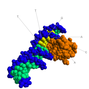
In Eukaryotic cells, a common promoter is a DNA sequence that reads TATAAA, and is better known as the TATA-Box. In bacteria, the common promoter is TATAAT, bettern known as the Pribnow Box (Pribnow-Schaller box). In both cases, the promoter is found in the Major Groove of the DNA Major Groovemolecule. As can be seen in the image to the right, the major groove is wide enough to "see" the base pairs. The base pairs have an electrochemical profile, and thus can respond to other chemicals (via van Der Waals forces). Thus, the major groove is a place where proteins (and other compounds) can bind to DNA! The promoter sequences are found in the major groove. Major Groove with Initation FactorThe image to the right shows a bacterial promoter event. One of the factors needed to start transcription (by recognizing the promoter) has bound into the major groove. This recognition event is needed to identify the start point of a gene. The Transcription Initation Complex will then begin to form at this site, and begin the transcription of the gene. 

Many genes are regulated, meaning they can be turned on and off. Beyond a promoter, a regulated gene will typically have a non-coding region known as the Operator. The operator is located down stream of the promoter (meaning it will be between the promoter and the coding region). Regulatory proteins can bind to the operator, preventing transcription. Remember, cells are masters at energy conservation. They will not begin producing proteins that are unnecessary. Gene regulation is a common activity of Signal & Receptor systems. The image below is a good visual of the promoter & operator systems. Gene House keeping genes are those that are needed for the general function of the cell, and can include genes for glycolysis, citric acid cycle, and ribosomes. These genes are always ON, and are referred to as constituative genes

Messanger RNA (mRNA) is a molecule of RNA that cares the gene code for the construction of a protein. mRNA is sent to the Ribosome in order to produce a protein. The code for constructing a protein is in Nucleotide Language, meaning the code is a code of nucleotides. Specifically, the code in mRNA is in the ribonucleotide language (A, U, G, C). In order to make a protein, it is necessary to Translate the ribonucleotide language into the language of proteins, i.e., amino acid sequcences. 

In order to translate, you need an agent of translation. This agent of translation must be a molecule that contains both ribonucleotides and amino acids (think of it as the nucleotide-amino acid dictionary). A specific ribonucleotide sequence must directly correspond to an amino acid, just as in translating human languages requires word for word relationships. This concept of a direct nucleotide to amino acid relationship is the basis of the Genetic Code.

tRNAThe agent of translation is Transfer RNA (tRNA). In tRNA, there is a direct physical correspondence between a 3 nucleotide sequence (anti-codon) and an amino acid. To the right are common ways of illustrating tRNA, with the 3rd image being the most common way of drawing the molecule. In the image, each molecule has a region known as the anticodon; this region will interact with mRNA. At the 3' end of the molecule, a specific amino acid will be bound. 

On the mRNA, the code is broken down into codons (think of these as genetic words). Codons consist of 3 adjacent nucleotides. Codons are complimentary to anticodons found on tRNA. Each tRNA has a specific anticodon-amino acid relationship, so each codon then specifies an amino acid. The genetic code is NOT ambiguous. There is a direct correspondence between codon and amino acid; the tRNAs make sure of this.
The ribonucleic language is divided into 64 3-nucleotide words known as codons. Condons specify though tRNA an amino acid. The Genetic Code is thus the translation scheme between codons and amino acids. [NOTE: another way to describe the genetic code is in terms of a computer algorithm]. Below is a rather unique way of viewing the genetic code. It is an excellent way of visualizing the number of redundancies in the code.
Genetic Code Algorithm
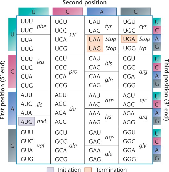
The genetic code is redundant, which means that there are multiple codons (3 nucleotides) that specify the same amino acid. For example, around the 12 o'clock position of the above chart, you see the amino acid glycine. The codons GGU, GGC, GGA and GGG all specify Glycine. Phenylalanine is specified by UUU and UUC. There are only a few amino acids, such as methionine, that are specified by a single codon (in the case of methionine it is AUG).

The presence of redundancies means that some alterations in the gene sequence are silenced (silent mutation). For example, changing GGU to GGA does not change the specified amino acid (Glycine). This is a silent mutation. Changing UUC to UUA may cause a problem (point mutation), but both Leucine and Phenylalanine are hydrophobic, so the variation may be minor. Chaing CAC to CAG though has more impact as you are changing the positive histidine to a polar glutamine (you loose the full positive charge of histidine). Remember, chaning amino acids can easily change the way a protein folds. REMEMBER: The genetic code has redundancies, and this will limit some problems with mutation.

Below is a more classic way to represent the genetic code, in the form of a table. The way the table is arranged, you can easily see the various redundancies in the system. In both representations, notice that there are three codons that specify STOP. These stop codons, UAA, UAG and UGA are essential for the termination of protein synthesis. In the image below, you will notice AUG has been tagged as the initiation (start) codon. All protein synthesis begins with the code AUG. We will talk more about this later in the the week.
Genetic Code

Daily Challenge

Your challenge today is just to discuss the concept of the Central Dogma. Don't worry about mechanisms yet. Consider the implication of the central dogma. In your own words, express why it is important. What are the important features? You can use questions above in "Today's Topic" as a way of focusing your thoughts.
Link to Forum

One final image

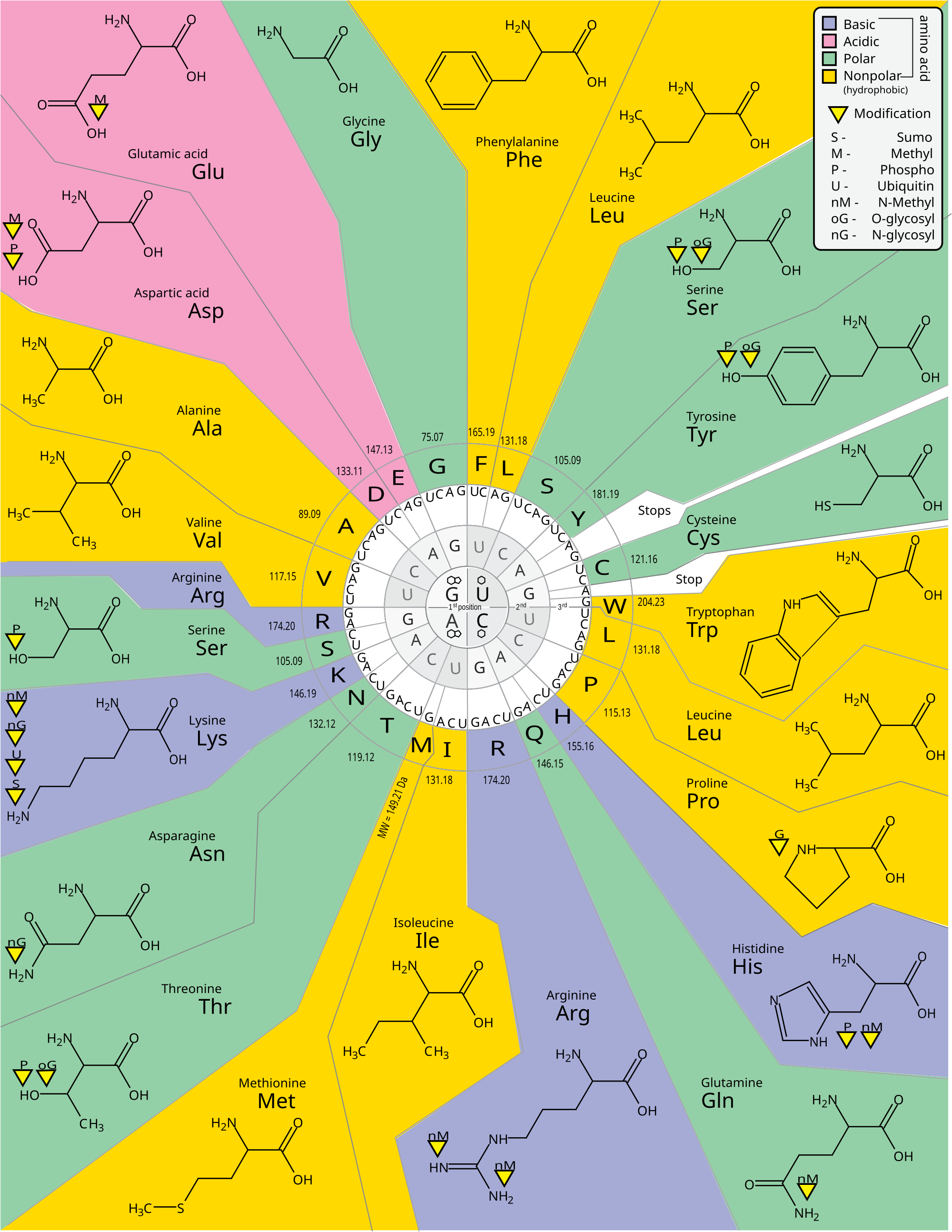
Below is a variation of the genetic code diagram shown above. In this image, the amino acids are drawn out and color coded by their type (basic, acidic, polar and nonpolar). Look over the chart. How easy is it to change amino acid type? If you change the first nucleotide in a codon, what is your chance of changing the amino acid type? What about the second codon? What about the third codon. An optional forum is open for you to explore these concepts.
Link to Forum
Genetic Code with Amino Acid Type

Friday, October 26, 2012

Daily Newsltter: October 26, 2012 - Mendel, Meiosis and Evolution

Site LogoDaily Newsltter

October 26, 2012

Mendel, Meiosis and Evolution


With meiosis we see the mechanism of inheritance first hinted at by Mendel. Gregor Mendel discussed how each person has two "copies" of the information that gives rise to a physical trait (genes), and that there were variations in that information which gave rise to different expressions of these traits (alleles).

Mendel's work gave biologists an understanding of inheritence, and an understanding of chromosomes and nuclear division helps to understand Mendel's inheritence model and the variations of that model. With cross-over in Prophase I, and the random seperation of homologs in Anaphase II, we can see the recombination of parental genes that result in genetically unique gametes (sperm and egg). This results in genetically unique individuals each generation.

Diversity is one of the greatest advantages of sexual reproduction. While >99% of our genes are held in common within a species, there is sufficent genetic variation to allow provide a species with a broad adaptaion range. Remember, the goal of evolution and survival resides not in the individual, but in the population. Those with genetic, metabolic, or physiological advantage will have a greater chance to leave offspring; thus the species survives and thrives.

In class, I mentioned that when you get to the core of biology, it is ultimately all about sex. Not the physical act, but the concept. The recombination of genes allows for diversity. Some organisms spend most of their life in haploid, or non-sexual reproductive states. They only expend the energy for sexual reproduction when they need to insure the survival of their offspring.

Daily Challenge


Today, you are to reflect upon what you have learned so far regarding metabolism, Mendelian inheritence, and meiosis. Build a picture of evolution based upon the need for a diverse population. Use an example to help illustrate the need for diversity. As a starting point, look to endangered species (why is it a problem when you have few individuals left?) or look at what happens when you have genetically similar trees struck by disease (Pine Bark Beetle infestations).
Link to Forum

Video Challenge


Today, I'm going to give you a video challenge. Below is a link to a video about animal genitalia (penises to be percise). The video is informative, but at times very vulgar (so be warned). The video's narrator makes some interesting, and humorous, comments about evolution.

What you are to do is reflect upon the views of evolution discussed. What insights can you glean from the narrator and the topic, and what errors are made (even jokingly) about evolution.

The World's Most Terrifying Penises: The Echidna

Link to Forum

Thursday, October 25, 2012

Special Edition: October 25, 2012 - DNA Replication

Site LogoSpecial Edition

October 25, 2012 DNA Replication


Textbooks have a tendency to make replication one of the most complext topics covered. With a tendency to throw all the current research and understanding at students, they rarely take a step back and try to explain it. This newsletters has two goals: 1) to help biology students understand DNA replication, and 2) to show you want is expected from a Biology Freshman/Sophmore.


Central Dogma of Biology
 Before we get into replication, let's take a step back and look at the three core genetic processes, a.k.a., the Central Dogma. The central dogma describes the flow of genetic information in a cell. The core idea is INFORMATION. You may recall some of our early discussion on DNA, and about base complimentarity and the directionality of the molecule. This will become important rather quickly, but for just this moment, I want you to concentrate on the fact that DNA carries information. Information on how to build RNA and Proteins, both of which will produce the phenotype (expression) of the cell. For this reason, DNA, RNA and Proteins are considered Informational Macromolecules. This means that the sequence of monomers contains infomation, e.g., instructions on how to build RNA and Proteins. Since it is critical, the sequence of nucleotides carries information.
As you can see from the diagram of central metabolism shown above, there are three processes: Replication, Transcription and Translation. Think about those words. They are words used in reference to languages and documents. When you replicate a docuement, you want to ensure that you are getting a faithful (or even exact) copy of the original.

When you transcribe, you are moving from one medium, e.g., spoken word, to another medium, e.g., text. If you watch news shows, they will tell you that transcripts of the show are available. Court reports make transcripts of the trial. You are taking the language from one medium (in our case DNA) to another medium (RNA). The language is still the same (i.e., nucleic acid lanugae), just in a different form. Does the transcript have to be 100% correct? You want it to be, but it is not as exacting as a replication.
Translation is where you change languages. Unless you're fluent in another language, you will need somoene to help translate, or at least a good translation dictionary. Now you are moving the context from one language (nucleic acid) to another language (amino acids).

All of these processes rely on one key feature of nucleic acids: BASE COMPLIMENTARITY. In DNA: A complements T, and G complements C. In RNA: U complements A, and G still complements C.

Replication

In replication our goal is to take one molecule of DNA and make two daughter molecules of DNA that are identical to the first. Even the best replication processes can produce errors, but our goal is to be error free. This takes precision! As DNA is long, we also need this to be a fast process.

The enzyme that is ultimately responsible for replication is DNA Polymerase. [NOTE: there are multiple types of DNA Polymerase, but for now you just need to understand the core concept common to the DNA Polymerase family.] DNA polymerase is only one component of the Replication Complex, which is a complex association of proteins needed to successfully complete the replication event. Your goal at this time is to concentrate on DNA polymerase; we will talk about some of the other components later.
DNADNA is a double stranded molecule, in which the strands are anti-parallel. This means that one strand starts at the 5' end 3' end, while the the other strand is revered. This can be seen in the image to the right. A common way of saying this is that we read DNA in the 5' 3' direction. Why is this important? DNA Polymerase can only read DNA in the 5' 3' direction, and can only build a new strand in the 3' 5' direction. Before we go on, let's look another time at how deoxyribonucleotides are polymerized.

DNA Polymerization
To the left you will see a generic image showing DNA Polymerase adding nucleotides to a growing DNA strand. Look carefully: A deoxyribonucleotide triphosphate (dTTP) is being added. The 5' phosphate of the new nucleotide is what will be used to form the phosphodiester bond. DNA Polymerase requires a free 3' end on which to grow the new DNA strand. To the right is another image that will help you with this concept.Phosphodiester Bond Formation At this point, the critical thing to remember is that DNA Polymerase will need a free 3' end on which to add a new nucleotide.
This requirement to build in only one direction (3' 5') creates a problem for the DNA process: the two strands read in opposite directions, and each must be replicated. It was noticed that one strand appears to replicate continuously, while the opposite strand appears to replicate discontinuously.

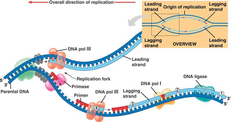
The original strand that reads 5' 3' can be used by DNA polymerase to continuously produce the new 3' 5' strand (the antiparallel complement to the original strand). We refer to the continuous synthesis strand as the Leading Strand. The other original strand, which read 3' 5' cannot be copied continuously. A section will have to be exposed, replicated, and then another section exposed. This strand is constructed discontinuously, and is reffered to as the Lagging Strand.DNA ReplicationThe image above shows the leading and lagging strand. Notice that the leading strand is replicating toward the Replication Fork(where the original strands seperate). As more DNA unwinds and opens, DNA synthesis continues down the leading strand.
The lagging strand though has to build in a start-stop action, producing Okazaki fragments. These fragments have to be sealed (phosphodiester bonds) together before DNA can rewind into the α-helix.

In the image above, you will notice a number of enzymes on the lagging strand. These enzymes are required for the initiation of DNA polymerazation, and then sealing the fragments. Looking at the image, you will see an enzyme called Primase, and a structure in red known as a Primer. Another restriction on DNA polymerase is that it must have a free 3' end from which to start building. DNA Polymerase is prevented from building a DNA strand from nothing. Something (a primer) must be in place upon which DNA polymerase can build. The Primer is constructed from RNA, and is a temporary scaffold upon which DNA polymerase can start working.

Eventually the primer will need to be moved. This is where you need to learn a little more about DNA polymerase. There are multiple DNA polymerases in eukaryotic systems. The general work horse of replication is DNA Polymerase III (DNA pol III), which is used to make long strands of DNA. DNA Polymerase I (DNA pol I) is used to replace primers (it is also used in repair functions). Even after the RNA primer is replaced with DNA, there is still a gap between fragments. Ligase is the enzyme used to create a phosphodiester bond between fragments, thus sealing the new sugar-phosphate backbone of the synthesized strand.

The result of DNA replication is that one molecule one DNA was used to create two new molecules of DNA. The two strands of the original DNA molecule became the template from which to build new complimentary strands of DNA. This is referred to as semi-conservative replication, as each new molecule has one strand from the original molecule, and one freshly synthesized complimentary strand.

Optional Challenge

In your own words describe the purpose and process of replication. While you are encouraged to deepen your knowledge of replication, you are only required at this time to go into as much detail as was presented in this newsletter.
Link to Forum

Link to Lecture Reflection Forum

Daily Newsletter: October 25, 2012 - Meiosis

Site LogoDaily Newsletter

October 25, 2012 Meiosis


On Tuesday, the topic discussed was mitosis. We saw that mitosis was standard nuclear division. When coupled with cytokinesis, we saw that the goal was taking one parental cell and creating two identical daughter cells. With meiosis, we will again experience nuclear division, but with a different goal than mitosis. The goal of meiosis is to produce gametic (reproductive) cells. As such, we will 'always' couple meiosis with cytokinesis.

Goal of Meiosis: From one diploid parental nuclei, generate four genetically unique haploid nuclei.

Goal of Meiosis-Cytokinesis: From one diploid parental cell, generate four genetically unique haploid cells.


The production of gametic cells requires two nuclear division events: a reduction division, and an equatorial division.
Important Note: All forms of nuclear division have the same basic four stages. Prophase is the set-up, Metaphase is when chromosomes align, Anaphase is when chromosome/chromatid seperate, and Telophase is when the nucleus returns to "norm".
The first meiotic divison is a reduction division, and is reffered to as Meiosis I. As the term reduction implies, we are "reducing" something. Specifically, during the reduction division the cell is reducing chromosomal number. REMEMBER: a diploid organism (2N) carries two copies of each chromosome. During a reduction division, the copies are separated and moved to opposite sides (poles) of the cell.

Homologous Chromosomes: This term refers to the two copies of each chromosome. For example: Chromosome 1 in humans is the largest of the chromosomes. Individuals have two copies of chromosome 1: Homologous chromosomesone from their mother and one from their father. These two individual examples of chromosome 1 are considered homologous. Homologous chromosomes carry the same genes, but each homolog (one copy) will have a unique set of alleles (one allele for each gene).
The image to the right is an example of of two different homologous chromosomes. Notice that at the same gene is found at same location on each homolog (a member of a homologous pair). As shown in the first homologous pair, one homolog carries allele A for the first labelled gene, while the second homolog carries allele a.

Homologous chromosomesIMPORTANT NOTE: Remember that what we are seeing are chomosomes. The two chromatids of a given chromosome are the products of DNA replication. The image to the left should help you remember that you start with a Maternal DNA molecule and a Paternal DNA molecule. After DNA replication, these will consense into maternal and paternal chrmosomes. It is critical that you remember the difference between Chromosome and Chromatid.

recombinationProphase 1 (prophase of Meiosis I) is the most critical stage of meiosis. Prior to the reduction division (which occurs in Anaphase 1), a recombination event will occur between the maternal and paternal chromosomes. Recombination is a genetic event in which homologous genes on two different DNA molecules are swapped between the molecules. A section of each DNA molecule is cleaved and then bound to a new molecule of DNA. The resulting molecules will still have the same genes, but the alleles they carried have been swapped.
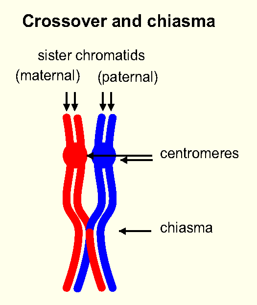
Crossover and ChiasmaThis recombination event begins when homologous chromosomes are brought together during prophase/prometaphase I. The homologs possess DNA sequence similarities, and are able to bind to each other (the homologs chemically recognize each other). Regions where Cross-Over (recombination) can occur begin to over lap, forming a visible structure known as a chiasma (pl. chiasmata). During cross-over(recombination), alleles are swapped between the two chromosomes, with the end result being genetically unique allele patterns on each chromosome. CRITICAL POINT: the result is two unique chromosomes! This step is critical for maintaining diversity in diploid (eukaryotic) cell systems. Ever generation inheriets a unique genetic composite of maternal and paternal alleles.
This event is one of the most critical ways that eukaryotic organisms ensure the diversity of their populations. Why?

After this recombination event, Chromosomes will line up the equitorial line (metaphase plate) so that there is one homolog on either side of the equitorial line. RECALL: in mitosis, chromosomes lined up so that there was a chromatid on either side of the metaphase plate; now in meiosis I, there is a homolog chromosome on either side. Why is this important?


metaphase
Metaphase in Mitosis
Meiosis I
Meiosis I


As you can see in the image of meiosis I, the alignment of chromosomes during Metaphase is critical. During anaphase I, the freshly recombined homologs are seperated. The cell moves from a diploid (having 2 sets of chromosomes, 2N), to a haploid state (having only 1 set of chromosomes, 1N). This is the reduction division!
Mitosis II occurs like mitosis, which is classified as an equitorial division. In an equitorial division, the chromatids are seperated. (see the difference?) Meiosis IINOTICE: each cell produced in meiosis I now undergo meiosis II.

During the reduction division (meiosis I), you went from a diploid to a haploid state. This is done by aligning the chromosomes in metaphase so that there is a homolog on either side of the metaphase plate. During anaphase, the homologs are seperated.

During the equitorial division (meiosis II), each chromosome is seperated into the individual chromatids. In metaphase II, chromosomes are aligned so that the chromatids are on either side of the metaphase plate (or to put it another way, they are alinged down the centromere). During anaphase II, the sister chromatids are then seperated.

The result: 4 genetically unique haploid cells!

Daily Challenge

We have discussed the main stages of nuclear division, and you have readings from your textbook and supplemental reading on meiosis. In your own words, describe the process of meiosis, complete with a discussion of synapsis, chisamata, and cross-over. Why is the recombination event so critical to population diversity? How could recombination affect evolution?
Link to Forum

Wednesday, October 24, 2012

Daily Newsletter: October 24, 2012 - The Cell Cycle

Site LogoDaily Newsletter

October 24, 2012 The Cell Cycle


Reading:

The Eukaryotic Cell Cycle
This is an excellent overview of the cell cycle. This is considered supplemental to what is in your textbook.


The cell cycle describes the life stages of a cell. It starts just after cytokinesis, and continues until the next cytokinetic (or mitotic) event. The period when a cell is growing between division events is known as interphase.

Cell CycleInterphase is divided into three distinct steps: G1 Phase, S Phase, and G2 Phase.

The G1 Phase stands for Gap 1 Phase, and begins just after cytokinesis. Gap refers to no visible change in the cell. Another good term for G is growth, and that is usually what happens during this phase. The cell is building primary products, compounds such as amino acids, phospholipids, carbohydrates, triglycerides. These are describe as primary products as they are the cellular biochemicals needed to increase the cells size (biomass). Thus we generally consider the G1 phase as the timethe cell increases in size. Another way to say this is that a major goal in this phase is the development of biomass.
[NOTE: In multicellular organisms, some cells will stop mitosis. They are locked in what is known as the G0 phase. The G0 phase is similar to the G1 Phase, but the cell will never level leave this physiological stage. After reading this newletter, I want you to consider a question: Why would you stop in the G0 Phase if you were never going to replicate again?]

When the cell gets a signal to divide, the physiology of the cell changes. Metabolic pathways for creating deoxyribonucleotides are unlocked (dATP, dTTP, dGTP, dCTP). The cell will use these deoxyribonucleotides to synthesize DNA (Replication). The production of enzymes needed to replicate DNA will also be begin. (Think back to cell signaling).

During the S Phase, cellular metabolism is focused on DNA replication (S stands for DNA Synthesis). This is a complex event, takes time, and has consequences if there is an error; so most of the cell's work is dealing with DNA replication.

Once S Phase has begun, the cell has committed its self to the process of Nuclear Division and Cytokinesis. You never replicate DNA without moving toward Nuclear division, DNA replication without nuclear division usually results in cell termination.

After DNA replication, the cell needs to build all of the proteins and compounds used during Nuclear Division and Cytokinesis. This is the goal of the G2 Phase. The G2 Phase will continue until the required components are constructed, and the cell receives the signal to continue. At this point the cell moves into the M phase (which stands for either mitosis or meiosis).


Regulation

The processes of the cell cycle is tightly regulated.
What happens in unregulated or uncontrolled cell growth?

The regulation is based on Signals (yes, we're back to signals). To maintain homeostasis, an organism must replace certain cells during its life time. For instance, humans replace skin and mucous membranes constantly. Hormones, such as insulin-like growth factors, are signals used to make sure the body maintains its self by replacing cells.
CDK
Inside of cells, there is an internal signal system based on the protein family Cyclin Depdendent Kinases (CDK). Note, this is a family of proteins involved in cell cycle regulation (they also have a few other functions). As the name implies, the protein needs a Cyclin to function. For example, CDK2 requires Cyclin E during G1. (NOTE: Cyclinn is a family of protein signal molecules assoiated with the cell cycle).

CDKs are always produced in mitotically active cells. (What term do we use for a protein that is always produced?)
Cells don't always have Cyclin, but instead produce them in response to a signal. [Note: The signals differ depending on if you are dealing with multicellular, colonial or singal celled eukaryotes, so we are not going to get into specifics. As these control vital processes that could cause damage to cells, e.g., think cancer, they have a very complex signal transduction. Think of it this way, the cell has to have get numerous "permissions" before it starts producing Cyclin, so it is highly regulated. For the purpose of this class, we will just say that it is controlled by "Growth Factors".]

CDK Check PointsThroughout the cell cycle, there are time points referred to as Check Points. These check points are where regulation occurs. For example, to move from the G1 Phase to the S phase, you need to produce a set of cyclins to induce the activity of CDKs. The Cyclin-CDK complexes can then phosphorylate proteins. Why would you need to phosphorylate proteins?

During the G1 Phase, the check point determines if you turn on the production of deoxyribonucleotides and the production of replication complex enzymes. There are check points in the G1 Phase, S Phase, G2 Phase, and M Phase. Many of these check points are determinations of the health of the cell, or the correctness of DNA replication and chromosome condensation. The image to the right shows the major check points, as well as the cyclin needed to activate (Cyc D to make deoxyribonucleic acids and the replication complex, Cyce to start the S phase, Cyc A to make sure replication is occuring properly, Cyc B to make sure the cell is ready for mitosis). Note: the check point in mitosis (M phase) is not shown. The M phase check point is to ensure that chromosomes have properly condensed and migrated.
As you can tell, the check points are there to make sure that the process is occuring properly. Consider the Cyclin/CDK system as the Quality Assurance & Quality Control (QA/QC) officer of the cell.

Go Phase (in case you missed it the first time around)
Some cells become non-mitotic at a given point in development. For example, nerve cells stop dividing, as do cardiac muscle cells. When a cell becomes non-mitotic, it shifts from the G1 Phase to the Go Phase. The cell can be locked from mitosis by blocking the genes for either CDK or Cyclin.

Daily Challenge

In your own words, describe the cell cycle. Then answer the following question: How are CDKs related to cancer?
Link to Forum

Optional Reading

If your really into the regulation of the cell cycle, try out this article:
Cyclins and Cell Cycle Regulation

Tuesday, October 23, 2012

Daily Newsletter: October 23, 2012 - Nuclear Division

Daily Newsletter

October 23, 2012 Nuclear Division


Mitosis describes nuclear division. We commonly use the term loosely to describe cell division, but that is an incorrect usage of the word mitosis. Cell division is better described by the term cytokinesis. It is important to use these terms correctly, as it will help you as you go further through biology.

Mitosis describes a nuclear division event. Most of the time we think of nuclear division occuring just prior to cytokinesis (cell division), but there are examples of cells that can undergo nuclear division without undergoing cytokinesis (many fungi do this). For most eukaryotic cells though, mitosis will be followed by cytokinesis. Remember though that these are two different events.

Mitosis is also described as somatic nuclear division. The word somatic refers to general body cells. This is to contrast difference between a general cell and a reproductive cell. A reproductive cell, or gametic cell, will undergo a special type of nuclear division known as meiosis, which will be discussed in Thursday's newsletter. Gametes are needed for sexual reproduction.

The goal of mitosis is to produce two daughter nuclei that are genetically identical. Coupling mitosis and cytokinesis results in the formation of two genetically identical daughter cells.

Prior to mitosis, DNA replication will have already occured. Every molecule of DNA will have undergone replication (DNA synthesis). We will discuss replication later this week.

The process of mitosis occurs in 4 Main Phases (there are additional phases that have been added in recent years). Below is a phrase to remember:

"Prophase sets up the process, metaphase aligns the chromosomes, anaphase seperates the chromosomes, and telophase returns the cell to normal."

These phrase describes the basic action of the four main steps: Prophase, Metaphase, Anaphase and Telophase.

[Review the University of Arazonia's Mitosis Tutorial. This will take you through the main phases, and includes the Prometaphase addition.]Prophase

In Prophase, we see the condensation of the chromosomes, which were discussed in yesterday's newsletter. We also see the formation of the mitotic spindle. The image to the right shows Early and Late Prophase. In recent years, the term Late Prophase has been renamed as Prometaphase. This was done to indicate the rapid changes that being to take place within the cell, and to indicate the passing of a check point (NOTE: cells go through check (or restriction) points where signals will tell the cell to either proceed or abort the process of nuclear or cellular division.

In Prometaphase, microtubules of the mitotic spindle reach toward the centromere of each chromosome, forming the kinetochore. During this period, we will also see the dissolution of the nuclear envelope. The nuclear lamella (intermediate filaments) begin to dissociate. As they do, the phospholipids of the nuclear membrane wrap around them forming submicroscopic vesicles (i.e.,can not be see under a light microscope).

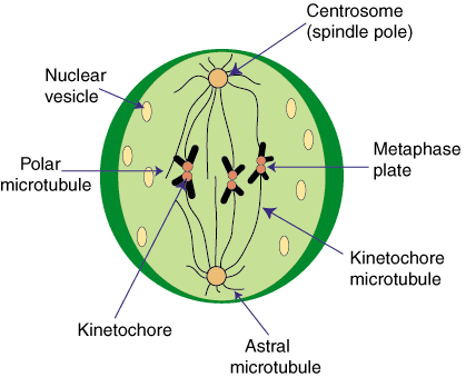
During Metaphase, the chromosomes are aligned down the imaginary equitorial line of the cell. This line is equidistant between the centrioles. The alignment is important. If you look at most drawings, the chromosomes are shown as aligning so that their centromeres are on the equitorial line, and individual chromatids are on either side of the line. This is to represent an equitorial division. As each sister chromatid of a chromosome represents a complete DNA molecule, the division of these will result in an equal number of chromosomes on either side of the equitorial line. So, a cell with 46 molecuels of DNA (chromosomes) will produce two dauther nuclei (and then cells) with 46 molecules of DNA (chromosomes). They will be genetically identical.

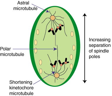
Beyond showing the arrangment of chromosomes, the image above also shows the different types of microtubules: Kinetochore, Polar and Aster. Aster, meaning star (Greek), comes from the starlike appearance of these microtubules. This starlike arrangment is also seen in the plant genus Aster, which is noted for the radial startlike flower petals. Note: they are sometimes referred to as Astral microtubules (Astral is an English adjetive derived from Aster).

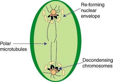
AnaphaseOnce the chromsomes have been arranged, they can be seperated. Anaphase is when there is visible seperation of the chromosomes into daughter chromatids. This link to an Anaphase Image is an excellent reference for what occurs in Anaphase (remember that you have motors that can move down microtubules). The image to the image to the right is a good quick visual of what starts to happen: the seperatation of chromatid. NOTE: At this point, there is no more chromosome; all we have left are the daughter chromatids. It is not uncommon though for people to start referring to these chomatids as chromosomes. As this can lead to a great deal of confusion, I want you to remember that at the end of anaphase we have daugter chromatids, not chromosomes.
Telophase
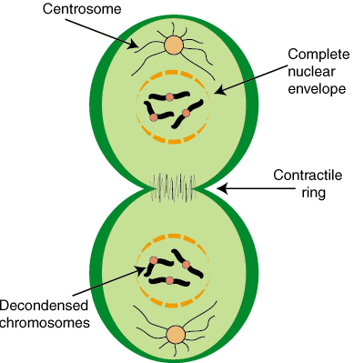
During Telophase, the cell returns to normal interphase operation. The kinetochore microtubules will be released and begin to dissociate. The nuclear envelope will reform as the nuclear lamella (intermediate filaments) begin to reassociate. Once the protection of the nuclear envelope is reestablished, the DNA will be released from the supercoiled packing that produced the chromatids and chromosomes. As this process continues, cytokinesis can begin.Cytokinesis
Remember, cytokinesis, or the division of the cell, does not have to take place after mitosis. For the vast majority of cells, mitosis and cytokinesis are coupled processes, but not all eukaryotes undergo cytokinesis. In animal cells, the act of cytokinesis is directed by the cytoskeleton, which causes the membrane to be pulled toward the center (remember the location of those polar microtubules?). As the membrane is dynamic, when pulled together, the phospholipid bilayers will "snap" together, or fuse, resulting in a seperation of the membranes.
Plant CytokinesisPlants and fungi, which posses cell walls, experience cytokinesis in a dramatically different way. Prior to division, the plant cell will have made numerous vesicles that contain the raw materials needed to create cell walls. These materials are held in an inert state, with inactive enzymes needed to form these materials into walls. As telophase begins, these vesicles begin to line up down the equator of the cell, and begin to fuse (what do you think triggers this action? will it trigger the enzymes?). As the vesicles fuse, cell walls begin to form. As more vesicles fuse (remember the membrane is dynamic), the wall continues to grow (cell plate). Eventually the membrane surrounded wall will fuse with the parental cell wall. Once the cell plate fuses with the parental wall, you have two new daughter cells.
PlasmodesmataNOTE: Plant cell walls are generally impervious to water flow. As a by-product of plant cytokinesis, and the development of the cell plate, you will find holes between the two new daughter cells. Known as plasmodesmata, these membrane lined holes allow for a continuous cytoplasm between cells. This allows for rapid movement of water and nutrients (including plant hormones) between cells (Symplastic Flow).


Study Note

You will notice that above I gave you a number of links. Do you think that they are important for your overall understanding of this topic?

Remember This: The following phrase will help you as we move through genetics. Remember it!

"Base complementarity is the foundation of all genetic processes."

Daily Challenge


Describe the process of mitosis in your own words. Feel free to use images, just reference where you got the image. Remember that this leads to your milestone paper and exam, spend some time to build a personal description of mitosis.
Link to Forum

Monday, October 22, 2012

Daily Newsletter: October 22, 2012 - Chromosomes

Daily Newsletter

October 22, 2012 Chromosomes


The word chromosome has a number of common means, but as biologists we need a strong (strict) definition of the word. For a biologist, a chromosome is a single molecule of DNA with associated packaging proteins, and is visible only during Metaphase and Anaphase of nuclear division. This second part is important; the chromosome is visible under light microscopy (the common form of microscopy). The chromosome is visible because it condenses (becomes tightly packed) during nuclear division (thus it is a eukaryotic phenomena).

We use the term chromosome to refer to a single molecule of DNA, but remember, in the strict sense, it only refers to a visible arrangement of DNA and proteins seen during nuclear division.

Another loose use is referring to the single, circular DNA molecule found in bacteria (or other prokaryotic DNA). As bacterial DNA is not held by the same packaging proteins, and has different packaging issues, the use of chromosome in this case is in error. The term genophore is a more appropriate term for prokaryotic DNA.

In eukaryotes, DNA must be packaged. One reason for this is to prevent physical strain or damage to the DNA molecule. The second reason is to conserve space. DNA is just too large when it is not packaged. The basic unit of DNA packaging is nucleosome. The nucleosome is a unit of packaged DNA, in which the DNA molecule is wrapped 1.67 times (~147 base pairs) around a core of 8 positively charged histone molecules (positive charge attracts negative phosphates).

These histone cores, when associated with Histone1 can begin to supercoil into a fiber. A cascade of interactions causes more supercoiling which ultimately forms the chromosome.
The chromosome when it first forms during prophase of nuclear division is actually two freshly replicated molecules of DNA (we just copied DNA). Below is a picture of an individual chromosome:
Notice that the two sides of the Chromosome are individual DNA molecules that we refer to as Chromatids. Holding the two chromatids together is a protein rich region known as the centromere. At the centromere, we will find a molecular motor known as the kinetochore. We will see the operation of the kinetochore during cell division.

NOTE: You have some words here that sound familiar and often get confused. In your notes, make sure you have definitions of Chromosome and Chromatid. Work on using these terms correctly.

Here is a video to help you visualize the process: How DNA is Packaged

Daily Challenge

In your own words, describe how DNA is packaged into a chromosome. Why is it important that we package DNA at the nucleosome and chromosome level?
Link to Forum

Other resources: