 Daily Newsletter
October 23, 2012 - Somatic Nuclear Division
Mitosis describes nuclear division of a somatic cell. We commonly use the term loosely to describe cell division, but that is an incorrect usage of the word mitosis. Cell division is better described by the term cytokinesis. It is important to use these terms correctly, as it will help you as you go further through biology.
Mitosis describes a nuclear division event, specifically in a somatic (as opposed to a sexual reproductive) cell. Most of the time we think of nuclear division occuring just prior to cytokinesis (cell division), but there are examples of cells that can undergo nuclear division without undergoing cytokinesis (coenocytic fungal cells have multiple nuclei enclosed by a single cellular membrane). For most eukaryotic cells though, mitosis will be followed by cytokinesis. Remember mitosis and cytokinesis are two different events.
Mitosis is also described as somatic nuclear division. The word somatic refers to general body cells. This is to contrast difference between a general cell and a reproductive cell. A reproductive cell, or gametic cell, will undergo a special type of nuclear division known as meiosis, which will be discussed in Thursdays's newsletter. Gametes are needed for sexual reproduction.
The goal of mitosis is to produce two daughter nuclei that are genetically identical. Coupling mitosis and cytokinesis results in the formation of two genetically identical daughter cells.
Prior to mitosis, DNA replication will have already occured. Every molecule of DNA will have undergone replication (DNA synthesis). We will discuss replication later this week.
The process of mitosis occurs in 4 Main Phases (there are additional phases that have been added in recent years). Below is a phrase to remember:
"Prophase sets up the process, metaphase aligns the chromosomes, anaphase seperates the chromosomes, and telophase returns the cell to normal."
These phrase describes the basic action of the four main steps: Prophase, Metaphase, Anaphase and Telophase. In the past ten years, prometaphase has been included as a stage to help distinguish events that occur as the cell moves from prophase to metaphase. You must remember that the stages are artificial attempts to describe a continuous process. Once the M Phase begins (in this case mitosis), it does not stop until completion or the cell receives a signal for apoptosis.
[Review the University of Arazonia's Mitosis Tutorial. This will take you through the main phases, and includes the Prometaphase addition.]

In Prophase, we see the condensation (packaging) of the chromosomes. We also see the formation of the mitotic spindle. The image to the right shows Early and Late Prophase. In recent years, the term Late Prophase has been renamed as Prometaphase. This was done to indicate the rapid changes that being to take place within the cell, and to indicate the passing of a check point (NOTE: cells go through check (or restriction) points where signals will tell the cell to either proceed or abort the process of nuclear or cellular division. This restriction point deals with whether the mitotic spindle is correctly forming or not (if not, apoptosis).
In Prometaphase, microtubules of the mitotic spindle reach toward the centromere of each chromosome, forming the kinetochore. During this period, we will also see the dissolution of the nuclear envelope. The nuclear lamina (intermediate filaments) and nuclear pores begin to dissociate.
During Metaphase, the chromosomes are aligned down the imaginary equitorial line of the cell. This line is equidistant between the centrioles. The alignment is important. If you look at most drawings, the chromosomes are shown as aligning so that their centromeres are on the equitorial line, and individual chromatids are on either side of the line. This is to represent an equitorial division. As each sister chromatid of a chromosome represents a complete DNA molecule, the division of these will result in an equal number of chromosomes on either side of the equitorial line. So, a cell with 46 molecuels of DNA (chromosomes) will produce two dauther nuclei (and then cells) with 46 molecules of DNA (chromosomes). They will be genetically identical.
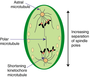
Beyond showing the arrangment of chromosomes, the image above also shows the different types of microtubules: Kinetochore, Polar and Aster. Aster, meaning star (Greek), comes from the starlike appearance of these microtubules. This starlike arrangment is also seen in the plant genus Aster, which is noted for the radial startlike flower petals. Note: they are sometimes referred to as Astral microtubules (Astral is an English adjetive derived from Aster).
 Once the chromsomes have been arranged, they can be seperated. Anaphase is when there is visible seperation of the chromosomes into daughter chromatids. This link to an Anaphase Image is an excellent reference for what occurs in Anaphase (remember that you have motors that can move down microtubules). The image to the image to the right is a good quick visual of what starts to happen: the seperatation of chromatid. NOTE: At this point, there is no more chromosome; all we have left are the daughter chromatids. It is not uncommon though for people to start referring to these chomatids as chromosomes. As this can lead to a great deal of confusion, I want you to remember that at the end of anaphase we have daugter chromatids, not chromosomes.
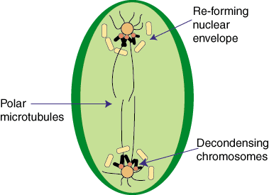
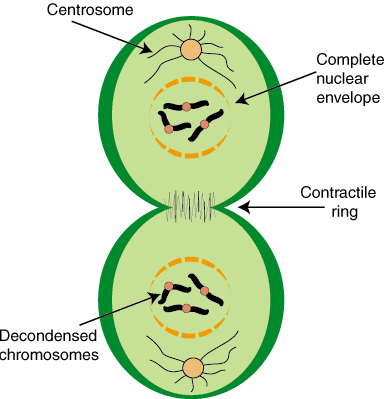
During Telophase, the cell returns to normal interphase operation. The kinetochore microtubules will be released and begin to dissociate. The nuclear envelope will reform as the nuclear lamella (intermediate filaments) begin to reassociate. Once the protection of the nuclear envelope is reestablished, the DNA will be released from the supercoiled packing that produced the chromatids and chromosomes. As this process continues, cytokinesis can begin.

Remember, cytokinesis, or the division of the cell, does not have to take place after mitosis. For the vast majority of cells, mitosis and cytokinesis are coupled processes, but not all eukaryotes undergo cytokinesis. In animal cells, the act of cytokinesis is directed by the cytoskeleton, which causes the membrane to be pulled toward the center (remember the location of those polar microtubules?). As the membrane is dynamic, when pulled together, the phospholipid bilayers will "snap" together, or fuse, resulting in a seperation of the membranes.

Plants and fungi, which posses cell walls, experience cytokinesis in a dramatically different way. Prior to division, the plant cell will have made numerous vesicles that contain the raw materials needed to create cell walls. These materials are held in an inert state, with inactive enzymes needed to form these materials into walls. As telophase begins, these vesicles begin to line up down the equator of the cell, and begin to fuse (what do you think triggers this action? will it trigger the enzymes?). As the vesicles fuse, cell walls begin to form. As more vesicles fuse (remember the membrane is dynamic), the wall continues to grow (cell plate). Eventually the membrane surrounded wall will fuse with the parental cell wall. Once the cell plate fuses with the parental wall, you have two new daughter cells.
 NOTE: Plant cell walls are generally impervious to water flow. As a by-product of plant cytokinesis, and the development of the cell plate, you will find holes between the two new daughter cells. Known as plasmodesmata, these membrane lined holes allow for a continuous cytoplasm between cells. This allows for rapid movement of water and nutrients (including plant hormones) between cells (Symplastic Flow).
Study Note
You will notice that above I gave you a number of links. Do you think that they are important for your overall understanding of this topic?Remember This: The following phrase will help you as we move through genetics. Remember it!
"Base complementarity is the foundation of all genetic processes."
Daily Challenge
Describe the process of mitosis in your own words. Feel free to use images, just reference where you got the image. Remember that this leads to your milestone paper and exam, spend some time to build a personal description of mitosis.

No comments:
Post a Comment