Friday, October 11, 2013

Daily Newsletter: October 11, 2013 - Light Dependent Reactions


Daily Newsletter

October 11, 2013 -  Light Dependent Reactions


We have looked at the harvesting of electrons by phototrophs (specifically in cyanobacteria and chloroplasts).  You will recall the image to the right. It shows the two photosystems, and the generation of NADPH and ATP.
Thylakoid Membrane
Recall the two photosystems are based upon pigment reaction centers.

The chloroplast of the the plant has two know Photosystems: P680 (Photosystem II) and P700 (Photosystem I).
  • Photosystem II
    • Used to Generate ATP (chemiosmosis)
    • Water is split to recharge the P680 reaction center with electrons and hydrogens.
    • Donates electrons to Photosystem I
  • Photosystem I
    • Can operate either Cyclic or Non-Cyclic
    • Non-Cyclic
      • Excited electrons are given to NADP+
      • Electrons accepted from Photosystem I to recharge the P700 reaction center.
    • Cyclic
      • Excited electrons are used to make ATP (chemiosmosis)
      • Electrons are returned to Photosystem I (recycled) to recharge P700 reaction center.
Remember: The photosystems are complex arrangements of chlorophyll pigments found in the thylakoid membrane. When electrons are used to make ATP, the electrons will be passed between electron carriers that act as proton pumps. The electrons are used to build and maintain a proton motive force, which will directly be used to make ATP. So all electron movement is occurring due to electron carriers in the membrane.
Now, what does all this mean for a plant? Throughout, we have talked about absorbance, but what is it? Simply put, absorbance describes the amount of light (photons) that is intercepted (absorbed) by the pigment. Each pigment has an absorbance spectrum, which is the wavelengths of light that the pigment can absorb.


Absorbance Spectrum Chlorophyll A and B To the right you will see the absorbance spectra of chlorphyll A and chlorophyll B. Both of these pigments are chlorophyll, thus we see them as green (but different shades of green). Each has a slightly different absorbance maximum; that would be the peaks on the graph (note the Y-axis is absorbance). So, Chlorophyll A absorbs strongly at 410nm, 430nm and 662nm, but best at 430 nm. Chlorophyll B absorbs best at 453. But there are more than just two pigments found in plants (and other photosynthetic organisms.

Plant Pigments AbsorptionThe graph to the left shows more pigements. Five pigments in all are shown. Notice that some, such as phycoerythrin and phyocyanin have absorption maxima at 550 and 610 respectively. Phycoerytrhin appears to the human eye as red (erythrin = red) and Phycocyanin is light blue (cyanin = blue). Carotene is characteristically orange. All of these, and more, can be found in plants. Why would a plant have so many different pigments?

To absorb as much light as possible. That is the #1 best answer!

Through evolution, plants have also mixed different pigments for other purposes, such as floral color attracting pollinators. For example, did you know that some flowers have UV pigments? Look at this image of the evening primrose. Evening PrimroseTo the left is what humans see, a full yellow flower; but under UV light, there is a large bullseye in the center. Insects see the bullseye, not the yellow flower. So pigments can be multipurpose.


Plants, and most other photosynthetic organisms, maintain multiple pigments to harvest as much light as they can. The ratio of pigments establishes the Action Spectrum of the plant, the wavelengths of light where they will have the highest yields of NADPH and ATP. This ratio of pigments will also produce distinctive colors. Are all plants green? Coleus Have you seen any shaded yellow? How about plants that have leaves with multiple colors? Here is a good example with Coleus. As you can see, there are different leaf coloration. Each variety would have different action spectrum, as each have a different pigment ratio. Why do you think plants would need different action spectrum? (Think survival strategy).

Consider a forest: You have a canopy of tall trees, then some intermediate sized trees and shrubs, then ground plants. Do shade plants have action spectra that differ from those of the canopy? Consider a plant that is being "shaded"; what does it mean to be "shaded"? You still get light, but is the light the same full spectrum light that the canopy is getting?

 
Going deeper into chloroplasts and the ancestral cyanobacteria, we find that there are distinct linages of chloroplasts.  Each lineage has different primary pigments, but you can find the biosynthetic pathways for all pigments in the cyanobacteria.  Below is a table published on the Wikipedia page for Chloroplasts.  The author of the image did an amazing job representing the various chloroplast lineages and demonstrating the pigments found in each lineage.  A clever point here is that the image author used the color reflected by the pigment (related to absorbance spectrum) and the organism (action spectrum) to provide a visual reference.  Make note that all the pigments can be found in cyanobacteria.


Daily Challenge

In your own words, discuss the concepts of absorbance spectrum and action spectrum. Show how they are related concepts, but describe different aspects of light absorption. Make sure you address the following questions:
  • Why do phototrophs need so many different pigments?
  • What does a single phototroph need more than one?
  • What is the difference between land plants and red algae that would have them being successful with different pigments?

Wednesday, October 9, 2013

Daily Newsletter: October 9, 2013 - Harvesting Photons

Daily Newsletter

October 9, 2013 Harvesting Photons


In photosynthesis, the cell will generate reducing power by using a photon to excite an electron. You will recall from chemistry that that electron excitation occurs when an electron jumps to a higher energy state (a higher shell).

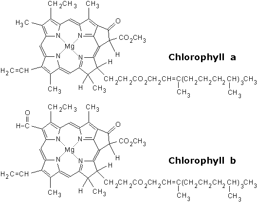
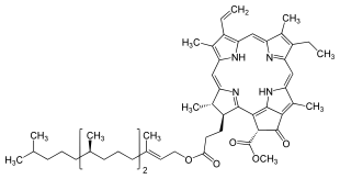
Photoexciation is one of the principle ways to excite an electron, and we can do this in a modern lab fairly easily. In nature though, you require a structure that can "focus" the photon's energy. Chlorophyll provides such a structure. As a pigment, the structure of chlorophyll is built to absorb specific wavelengths of light, and then excite a set of electrons.
Note that at the center of the chlorin ring (the organic ring structure) is a Magnesium ion, which can be considered the focal point of the photoexciation. Remember our discussions about the respiratory chain, specifically NADH dehydrogenase? Fe-S complexes were used for redox reactions with the protein. Magnesium, like iron, can easily go through redox reactions. As a point of reference, visualize the photon exciting the magnesium ion.

Chlorphylls are arranged in systems, and once photoexcitation has occurred, the energy is transferred by resonant energy transfer to a Reaction Center.
Resonant energy transfer? What's that?
This is a concept from quantum physics. It describes how energy is transferred between chromophores (e.g., Chlorophyll). One chromophore is struck by a photon and is excited. Instead of giving off light (i.e., fluorescence), the chromophore transfers the energy to a neighboring chromophore (if you are curious, this occurs through dipole-dipole coupling). As with any energy transfer, the 2nd law of thermodynamics applies. The amount of energy lost is inversely proportional to the distance between chromophores (it is very energy efficient between close neighbors). The important thing to remember here: we don't move electrons, we just transfer energy. This is possible due to the structure of the chromophores (i.e., pigments).

Energy is passed between chromophores in an antennae complex toward a reaction center. The reaction center is formed by a pair of chlorophyll molecules, and is chemically active, i.e., the reaction center can undergo redox reactions. There are two specific reaction centers known in plants, the P680 and P700. These names refer to the maximum red absorbance wavelength for these two molecules. The P680 reaction center is found Photosystem II, while P 700 is found in Photosystem I. (Photosystem is the name given to a reaction center and the associated electron transport chain. The naming is historic: Photosystem I was found before Photosystem II).


The reaction centers gather the energy absorbed by the surrounding chlorophyll. These reaction centers then undergo a charge separation, which is a form of redox reaction that donates high energy electrons to a waiting quinone. The image below is of photosystem II.

There are a few important points in this image:
1) Notice in the center there is a group f structures with the code Pheo. This represents a Phenophytin, which is a chlorophyll without the magnesium in the center.
The paired chlorophylls become a paired phenophytin. This change (loss of the magnesium) is a where the absorbed energy is converted to a redox potential. The energy is handed off to a quinone (Q).
You will recall ubiquinone, which we saw in the eukaryotic respiratory chain. Ubiquinone was a type of quinone. So this is a hydropobic mobile electron carrier (i.e., it can travel in the lipid portion of a phospholipid bilayer). The quinones take the harvested reducing potential to electron carriers for processing.

2) Notice that the reduction of the quinone requires Fe. Recall that Fe-S complexes were used in the respiratory chain for redox reactions. Iron is again used to help faciliate redox reactions.

3) Photosystem II has a way to recharge the system with electrons and hydrogen. The reaction center of photosystem II has hydrolytic ability; it can break water. The hydrogens, and their electrons, join photosystem II, recharging the reaction center (they convert pheophytin back to chlorophyll). In the process, they release oxygen (O2).

This is critical: the individual pigments do not undergo redox reaction; instead they transfer energy to neighboring chromophores (i.e., pigments). The reaction center undergoes redox reactions, passing electrons to ubiquinone. The reaction center lost electrons, and must replace these electrons. Photosystem II handles this replacement by using H2O. (Connection: H2O was created due to the reduction of the final electron acceptor O2. Now H2O is used to replace the missing electrons in the P680 reaction center, and O2 is given off).

The harvested electrons will be used to either make ATP (chemiosmosis) or to reduce NADP+ to NADPH + H+.

NOTE: Photosynthesis uses Nicotinamide Adenine Dnucolotide Phosphate. NAD+ is used in catabolic reactions, while NADP+ is found in anabolic reactions.

Photosystem II & I - Non-cyclic

Photosystem II

Daily Challenge

Explain in your own words how photon energy is harvested and converted into reducing power. Include in your discussion a description of the above electron transport system involved in producing NADPH + H+ and ATP.

Tuesday, October 8, 2013

Daily Newsletter: October 8, 2013 - Carbon Fixation & Calvin Cycle

Daily Newsletter

October 8, 2013 Carbon Fixation & Calvin Cycle


Our first stop in understanding photosynthesis is to first look at the autotrophic aspect, i.e., how we take Fix CO2 into organic carbon, and then reduce the resulting carbon compounds to produce glucose (Calvin Cycle).

Calvin CycleThe Calvin Cycle is a complex set of reaction that can be broken down into three main stages: Carbon Fixation, Reduction, Regeneration. The diagram to the right is a very minimized cartoon of the Calvin Cycle, provided only to acquaint you with the three stages.

One of the most important reactions in biology is the carbon fixation step of the Calvin Cycle. This is not hyperbole. This one stage of the Calvin cycle is the major way that CO2 is maid available to living systems. CO2 is a highly oxidized compound, and is considered an inorganic compound. It is also very stable. We have to some how take this compound and add it to an organic structure, thereby making it available to other living systems. This is the critical step in autotrophy.

[NOTE: Some bacteria can use other biochemical pathways for carbon fixation, but the vast majority of carbon fixation will occur in reactions similar to that which occurs in the Calvin Cycle.]

Consider what it means to have bioavilable carbon (i.e., carbon usable by non-autotrophic organisms). Consider what you ate today? Did it consist of starches, vegetables, animal products? Did it have protein, carbohydrates, lipids? All of these are organic (biochemical) molecules. They are all made of carbon, and we can consider them derivatives of Carbon Fixation. You may have heard that all of our energy comes from the sun; well the carbon fixation reaction is where we get the carbon to hold that energy (i.e., reducing power).
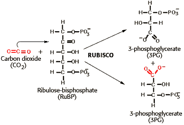
Above is a great diagram of the carbon fixation step in the Calvin Cycle. CO2 is added to the second carbon (carboxylation). There is an unstable intermediate that is formed, which spontaneously splits into two molecules of 3-Phosphoglycerate (3GP).

Ribulose 1,5 Bisphosphate Carboxylase (RuBisCo) is the enzyme that catalyzes the carbon fixation step. This enzyme is often cited as the most abundant enzyme on the planet, which is most like the case. No one though could doubt that it is the single most important enzyme, for without this, we would not have the quantity of organic carbon that is needed to support life as we know it.

Carbon Cycle

One area of study in ecosystem ecology is the cycling of nutrients through the environment, and one of the most critical cycles is the Carbon Cycle. The diagram below is a simple depiction of the carbon cycle, and Carbon Cycleshows the routes that carbon can go through between biotic (living) and abiotic (non-living) systems. The focus is to look at the CO2 cycle in the diagram. CO2 is incorporated into plants through photosynthesis. Animals eat the plants, extract energy and carbon, and release CO2 (Glycolysis & Citric Acid Cycle). Plants also respire (they have mitochondria), and decay organisms (bacteria and fungi) also respire. This is the most dynamic aspect of the carbon cycle.
Is there other carbon? Yes! There are fossil fuels, limestone deposits, and other geological carbon sinks. But it is the relationship between photosynthesis and respiration that is the strong and dynamic feature of the carbon cycle. The step of Carbon Fixation is the critical reaction.

RuBisCo

Ribulose 1,5 Bisphosphate Carboxylase is a fascinating enzyme, and has been the subject of a great deal of research. As the name implies, the substrate is a five carbon sugar (ribulose) that contains two phosphate, bound to the 1st and 5th carbon respectively. It's action is to add a carboxyl (CO2) group to the ribulose 1,5-bisphosphate.

It is also an enzyme that can carry out the reverse reaction, and can do so quite easily (which is a problem). This is one of the few enzymes where equilibrium becomes important. There is a problem known as photorespiration where RuBisCo reverses, and starts releasing CO2.

Calvin Cycle

Remember: we can break the Calvin Cycle into three stages: Carbon Fixation, Reduction and Regeneration. Calvin Cycle

The diagram to the right gives a little more detail on these stages. Above, we discussed the carboxylation of ribulose 1,5-bisphosphate (Carbon Fixation).   We must now look at the rest of the cycle.  

The product of the carbon fixation reaction, Phosphoglycerate (PGA) is the the substrate for the reduction stage of the Calvin cycle. As the diagram shows, this is where ATP and NADPH + H+ is used. The reduction stage ends with the production of Glyceraldehyde 3-P (G3P). Two out of every 12 molecules of G3P created will be used to make a hexose (e.g. glucose). The other 10 molecules of G3P will be used to regenerate the 5 carbon RuBP. The conversion of G3P to RuBP is the regeneration step.

Note that the calvin cycle must run multiple times to get a single glucose molecule. 6 RuBP must be carboxylated. 12 PGA must be reduced. 10 G3P must be regenerated. It is not as simple as glycolysis and citric acid.  Consider, you have to take 10 three-carbon structures, and convert them into 6 five-carbon structures during the regeneration steps; how do you do that?

The following diagram gives you some of the complexity of the Calvin cycle. You are not required to memorize this, but with your growing knowledge of reactions, you may find you can follow it.

If you notice, the set of reactions at the top of the image show the Carbon Fixation and Reduction steps. Everything else deals with regeneration. Recall that with glycolysis and citric acid, the intermediates were used for other reactions. The same thing is happening here. These intermediates of the regeneration stage can be used for other chemical processes.
Calvin Benson Cycle

Daily Challenge

In your own words, discuss the connection between this step and the events of glycolysis and the citric acid cycle. Where did decarboxylation take place? Why is RuBisCo and the carbon fixation reaction so critical? How would you turn Glyceraldehyde 3-P formed in the Calvin cycle into glucose?  Can you figure out the reactions in the regeneration steps?  Why did we need NADPH?
Link to Forum

Monday, October 7, 2013

Daily Newsletter: October 07, 2013 - Introduction to Photosynthesis

Daily Newsletter

October 07, 2013 Introduction to Photosynthesis


For this discussion, we will focus on Photosynthesis in eukaryotes, specifically in the chloroplast.

Chloroplast

Like the mitochondria, the chloroplast is an endosymbiont.  The cyanobacteria (photosynthetic bacteria) is the prokaryotic relative of the modern chloroplast.  The cyanobacteria became an endosymbiont after the formation of the mitochondria (remember, all eukaryotes carry mitochondria (or at least mitochondrial DNA), but only photosynthetic eukaryotes have chloroplasts).  There are different lineages of chloroplasts, just as there are different lineages of mitochondria.  NOTE:  Cyanobacteria is a phyllum of bacteria.  The chloroplast is considered a part of that phyllum.

Figure 1: The Chloroplast - http://en.wikipedia.org/wiki/File:Chloroplast_II.svg
The structure of the chloroplast has been maximized for photosynthesis. As can be seen in the image to the right (figure 1), you have an organelle that is composed of multiple membrane compartments.

The outer chloroplast membrane is eukaryotic in nature, while the inner membrane is bacterial in origin and composition.  In some plants, there is even a peptidoglycan cell wall (bacterial cell wall) between the outer and inner membrane.

Inside of the inner membrane, the chloroplast can be divided into the Stroma and the Thylakoid Membranes.

The stroma is the fluid filled inner compartment of the chloroplast, while the thylakoid membrane is an internal membrane that specializes in the harvesting light (photons) and converting the energy into reducing power.The thylakoid membrane is divided into two main types:  Stromal Thylakoids (aka Lamellae or Frets) and Granal Thylakoids.  The granal yhylakoids make up the stacks of thylakoids known as Granum, while the stromal thylakoids create supporting structure.  The entire system of thylakoid membranes is suspended inside of the stroma.

The thylakoid membrane system has its origin in the Cyanobacteria (see figure 2).  The cyanobacteria are one of the bacterial groups able to create internal membranes.  They form Photosynthetic Lamellae, which are the precursors to the thylakoid membrane. This lamellae provides an increased surface area for pigments (phycobilisomes).  This increases the chance that a photon will strike a pigment in the correct orientation.  If it were not for the increased surface area, the bacterium would have very little energy to carry out carbon fixation.  (Remember:  To get enough energy, a phototroph needs an increased surface area).

Figure 2: Cross-Section Structure of Cyanobacteria-http://images.tutorvista.com/content/kingdoms-living-world/cyanobacteria-cell-structure.jpeg

Photosynthesis

Photosynthesis is divided into two stages:  the Light Dependent Reactions and the Calvin Cycle (aka Light Independent or Dark reactions).

The light dependent reactions involve the pigments (photosystems) and proteins of the thylakoid membranes.  Within the system of thylakoid membranes, NADPH + H+ and ATP will be produced.  You will see the formation of a proton motor force, which will be used to make ATP.  Note that we are not using NAD+, but instead NADP+.  As a generality, NAD+is the electron carrier used in catabolic reactions, while NADP+ is the electron carrier used in anabolic reactions (why?  does it have to do with the enzymes being used?).

The energy (reducing power and ATP) created during the Light Dependent Reactions will be used in the Calvin Cycle to reduce carbon compounds, with the end result being the production of glucose. In your studies of biology, you may have seen the following reactions:
Cellular Respiration
C6H12O6 (s) + 6 O2 (g) → 6 CO2 (g) + 6 H2O (l) + heat
Photosynthesis
light + 6CO2 + 12H20 --> C6H12O6 + 6O2 + 6H20 

They look simple, but as we have discussed with cellular respiration, it takes many steps to get to the final product. Likewise, with photosynthesis, we are not going to complete this general reaction in one step. One common misconception is that Cellular Respiration is the reverse of Photosynthesis. On the surface they may appear to be reversals, but you're not going to see cellular respiration solely in reverse (there are times that you will see some reactions from glycolysis).  


Daily Challenge 

Compare the origin, structure and function of the chloroplast and mitochondria. The mitochondria had a folded inner membrane, and in the chloroplast, there is an entire system of internal membranes. Why is surface area so critical to the functions of these organelles?  Why do critical reactions (Calvin Cycle and Citric Acid Cycle) take place within these organelles?  If we but chloroplasts into a human, could they become photoautotropic?

Thursday, October 3, 2013

Daily Newsletter: October 3, 2013 - Respiratory Chain & Fermentation

Daily Newsletter

October 3, 2013 Respiratory Chain & Fermentation


The metabolic pathways of glycolysis, formation of acetyl CoA, and the citric acid cycle provide the means of harvesting reducing potential from the complete breakdown of glucose. While there are a few substrate level phosphorylation events, the bulk of energy harvested is in the form of reducing potential held by reduced electron carriers. But what happens to these electron carriers.

As with other aspects of metabolism, they can have multiple uses. For example, some biosynthesis reactions will require reducing potential in the form of reduced electron carriers. The bulk of the electron carriers though will be used to power respiratory chains.

In prokaryotes, the respiratory chain will be found on the cell membrane. These respiratory chains will use the reducing potential to power cell membrane activities, such as ion pumps (active transport) and flagellar spinning. They will also use their respiratory chain to power the production of ATP.
The vast majority of eukaryotes, in contrast, do not have repiratory chains embedded in their membrane. Instead, they rely on their mitochondria. Recall that the inner membrane of the mitochondria is prokaryotic in origin. The mitochondria specializes in using reducing potential to power the production of ATP; eukaryotes mitochondria & respiratory chainin turn utilize ATP for enzyme activation and activity. The mitochondrial inner membrane is covered in the proteins related to respiratory chains, and this specialization makes them more effective at using reducing potential than their prokaryotic relatives.

Overview

The respiratory chain (aka, the electron transport chain), is found embedded in the inner mitochondrial membrane. These membrane proteins function as electron carriers, but unlike NAD+ and FAD, they are enzymes that utilize reducing potential to pump H+ from the mitochondrial matrix across the inner membrane into the intramembranal space.

Therefore, we have reducing potential providing the energy to pump H+; or put another way, primary active transport (directly using energy to pump). The intramembranal space is small, and is being filled with H+, so this region will be acidic. We are also creating a very strong H+ electrochemical gradient across the inner mitochondrial membrane (potential energy). This gradient is called the proton motive force.

Hydrogen will move though open H+ channels back into the matrix (facilitated diffusion). When an ion moves down its electrochemical gradient, across a membrane, work is done (kinetic energy). The mitochondria utlizes this kinetic energy by having the H+ channel complexed with enzymes that make ATP. As H+ comes back into the matrix, the kinetic energy released though the ion's movment is used to make (regenerate) ATP. This is known as the Chemiosmotic Theory (movement of H+ down its electrochemical gradient across a membrane powers ATP synthesis).

Specifics

The electron carriers reduced during the citric acid cycle (which occurs in the mitochondrial matrix) go to NADH Dehyrogenase, also known as complex I of the respiratory (electron transport) chain. The electron pair will be passed to NADH dehydrogenase, NADH Dehydrogenasewhile the 2H+ will be pumped across the membrane into the intramembranal space. The image to the right shows our current understanding of NADH Dehydrogenase. NADH+ gives the electrons (reducing potential) to a flavin group (FMN). The electrons are subsequently passed through 8 Iron-Sulfar complexes. It is in these sequential redox reactions that the protein changes shape, allowing hydrogen to be pumped into the intramembranal space. The electrons are then passed to a mobile electron carrier known as ubiquinone, which has the ability to move around inside a phospholipid bilayer.

Why is all of this important? Well, why do you need iron in your diet? Why do organisms that have respiratory chains need iron? The main reason is that iron is an excellent reactant in redox reactions. Iron readily moves through different redox states, so is a great mineral to use when trying to move electrons. We also need iron for oxygen transport in the blood, but cells need iron to move electrons. You will hear more about iron-sulfur complexes in higher level classes.

The flavin group is a strong electron acceptor, and makes NADH a flavoprotein. Flavin groups in proteins can have odd reactions, one of the most critical is the donation of an unpaired electron to O2, forming potent reactive oxygen species such as Superoxide (an oxygen free radical) and the peroxide free radical (hydrogen peroxide). Reactive oxygen species have an unpaired electron, and they become potent oxidation agents (they pull electrons from other molecules). They have the ability to disrupt all organic compounds, including biopolymers. The peroxisome (a vesicle microbody found in cells) is responsible for clearing the cell of reactive oxygen species. NOTE: the formation of the free radicals is a natural by-product of flavoproteins such as NADH reductase.

Ubiquinone is important as an intermediate in electron chains. Ubiqunones can move around the inner membrane, connect the stationary transmembranal H+ pumping electrons carriers. In prokaryotes, the quinones act as a pool, and can accept electrons from sources other than NADH. This allows some procaryotes to harvest electrons from reduced minerals (more about that in microbiology).

NOTE: At this academic level, I want you to become familiar with NADH reductase as an example of a transmembranal H+ pump and electron carrier. The rest of the complexes we will cover in more superficial details.

Ubiquinone will then pass the electrons to complex 3, Coenzyme Q-Cytochrome C Reductase. This transmembranal protein accepts electrons from ubiquinone (which is also known as Coenzyme Q, hence the enzyme name). The protein will use the reducing potential to pump H+ into the intramembranal space. The electrons will then be given to Cytochrome C (cytochrome C will be reduced). NOTE: Coenzyme Q (ubiquinone) is also part of the cells defense against free radicals.

Coenzyme Q is considered an Anti-oxidant (due to its redox capabilities). Since ubiquinone is able to move in the lipid portion of the bilayer, it is lipid soluble. In digestion, it is absorbed through the same route as lipids. Can takinig a Coenzyme Q supplement help me? There are some conditions (like Huntington's disease) where Coenzyme Q is recommended, but there is not enough robust clinical data to confirm benefits in other conditions or athletic performance. Do your research before taking supplements. (BTW...what is implied by the word robust in the above sentence?)

Cytochrome C is another mobile electron carrier, but in this case it is water soluble and must move along the surface of the membrane. This protein is contains a Heme group (like in hemoglobin), which carries single electrons. In prokaryotes, Cytochrome C has been shown to have detoxification properties (oxidizing or reducing compounds), and in eukaryotes it plays a part in apoptosis.

Cytochrome C donates electrons to Complex 4, Cytochrome C Oxidase. This is the final transmembranal H+ pump, and the reducing potential is nearly spent in this pumping action. Cytochrome C Oxidase passes these "spent" electrons to oxygen, forming water. Oxygen is thus the FINAL ELECTRON ACCEPTOR. One interesting aspect about Complex 4 is that it uses copper (Cu) for redox reactions.
What about complex 2?

There is a complex 2, but it is not involved when NADH+ + H+ is used. Complex 2, aka Succinate Dehydrogenase, accepts electrons from FADH2, and it is not transmembranal (so no H+ pumping action). This is an interesting enzyme ☺. Do you remember the newletter yesterday about the citric acid cycle? The conversion of succinate to fumarate is handled by Succinate Dehydrogenase! This is the same enzyme! This is another example of the differences between isolated reactions and reactions inside of cells. For the longest time, chemists thought that FAD would have to be an intermediate in the movement of electrons. Modern biochemistry though has shown that the enzyme that handles the oxidation of succinate is part of a complex of enzymes (quantrenary protein structure) that is known as complex II. In the image of the citric acid cycle shown yesterday, the image author used Q instead of FAD to represent the electron carrier. The reason should now be clear...(hint: succinate is oxidazed, and the electrons are used to reduce ubiquinones. The important thing to remember is that the reducing potential that enters here has missed complex I, NADH Dehydrogenase, which is the first H+ pump. This means that reducing potential is only usuable by Complex III and IV.

Each of the transmembranal H+ pumps will move H+ from the matrix to the intramembranal space. This produces a high H+ electrochemical gradient (potential Energy). ATP synthase is another transmembranal protein found in the inner membrane. It acts as a H+ channel (facilitated diffusion). As H+ moves through the channel, toward the matrix, the enzyme is able to produce ATP from ADP and Pi. Since the reducing potential of NADH+ + H+ was used in complex I, III, and IV, it produces more ATP than the reducing potential harvested by complex II (succinate dehydrogenase).
For our purpose, we will round up the actual figures to whole numbers. NADH+ + H+ will allow for the regeneration (synthesis) of 3 ATP, while the reducing potential harvested at Complex II (or historically, FADH2) will yeild 2 ATP. NOTE: These are actually over estimates, but they work to simplify the math.


ATP Totals

What follows is a discussion of the Theoretical Yield of ATP from the catabolism of 1 molecule of Glucose. Note the phrase theoretical yield. Remember that cells have to decide between energy and carbon precursors, so cells rarely make it this high. We are also using whole number estimates of ATP production. An actual cell will have lower ATP yields. Also remember, eukaryotic mitochondria are more efficient than prokaryotes, and prokaryotes have other uses for the proton motive force. Eukaryotic actual yeilds will be higher. These numbers are also for ONE molecule of Glucose.
Glycolysis
  • 2 ATP used to phosphorylate intermediates
  • 4 ATP generated by Substrate Level Phosphorylation
  • 2 NAD reduced to NADH+ + H+.
    • NOTE: The NADH+ + H+ gained from glycolysis will only produce 2 ATP each. It is the cost of being reduced in the cytoplasm and then having electrons passed to the inner mitochondrial membrane.
    • So, the 2NADH+ + H+ will yeild 4 ATP.
Formation of Acetyl CoA
  • 2 CO2 released.
  • 2 NAD reduced to NADH+ + H+.
    • through respiratory chain → 6 ATP
Citric Acid Cycle
  • 4 CO2 released.
  • 2 GTP → 2 ATP generated by substrate level phosphorylation
  • 6 NAD reduced to NADH+ + H+.
    • through respiratory chain → 18 ATP.
  • 2 QH2 (FADH2)
    • through respiratory chain → 4 ATP.
Theoretical NET ATP generated: 36 ATP
Where did most of the energy come from? Reducing Potential
Where was most of the ATP made and how? ATP synthase, using the proton motive force generated by the respiratory chain.
What metabolic pathway has the highest ATP yield? Citric Acid Cycle.

Fermentation

What happens if the mitochondria runs out of oxygen?

Remember, oxygen is the final electron acceptor. Once the electrons have powered the H+ pumps in the inner mitochondrial membrane, they are at low energy. Electrons don't disappear, they have to be given to another atom/molecule. With the mitochondira, Complex IV passes the electrons (and some hydrogen) to Oxygen, thus forming water.
If not replenished, oxygen concentrations in the mitochondria will start to drop. For humans, this will happen when we either can not get enough oxygen through breathing or when our cells are metabolically very active (e.g., a work-out). If we don't have oxygen, then we don't have a final electron acceptor.

When there is no electron carrier, then Complex IV can not get rid of the electrons it holds. It can not accept electrons from Cytochrom C. If Cytochrom C can not reduce something, then it can not accept electrons from Complex III. This means Ubiquinone will not be able to reduce Complex III, and so can't accept electrons from Complex I. Ultimately, this means that complex I can not oxidize NADH + H+.

Is that really a big deal? YES. Remember talking about Flavin and how it can act up, creating free radicals? One trigger for creating free radicals is an imbalance in the expected ratio of NAD+ and NADH + H+. When you get too much NADH + H+ Complex I starts forming free radicals. (NOTE: this is only one outcome...the point is we need to regenerate a quality of NAD+)

When eukaryotic cells start running out of NAD+, Pyruvate Dehydrogenase becomes inactive (think regulation). This means that the cell has to do something to the building concentration of pyruvate (remember...we don't want to build up product...product should always become a new substrate). Pyruvate is moves to a different reaction or pathway.

At this point remember that the trigger for the shift is a loss of NAD+ and a build up of NADH + H+.

In animal cells, protist, and many fungal cells, the pyruvate will be oxidized into Lactate. In some fungi, the pyruvate enters a small pathway that oxidizes and decarboxylates the molecule, resulting in Ethanol (alcohol fermentation). fermenation
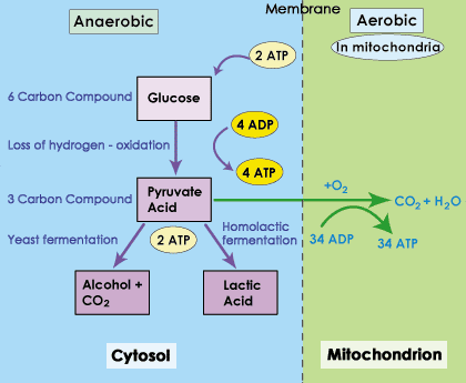
So, what is the result of moving to fermentation. In fermentation, we have to oxidize pyruvate. Electron carriers are needed for redox reactions, so in the oxidation of pyruvate NADH + H+→NAD+. This regenerates NAD+. Since we have NAD+, we can also carry out the substrate level phosphorylations that occur in Glycolysis.


You will recall that the substrate level phosphorylations of glycolysis are not strong energy harvests. More energy is harvested in redox reactions than in substrate level phosphorylation. So, why does it matter that we have some minimal ATP production?
Your cells have critical enzymes that have to keep working for the cell to survive. In an anaerobic state (no oxygen), you don't have enough ATP to carry out all life functions, but there is enough thanks to fermentation to keep critical enzymes running. The goal is then to bring more oxygen into the cells.

Is there a time limit? YES, but it depends on the cell in question. Some fungi can survive in fermentation indefinitely (but they may not be able to survive they're by-product, e.g. alcohol). For other cells, yes, there is a limit. Lactate in water is Lactic Acid, and it does alter pH. More importantly, there are some cells that have more critical function than others. Neurons in the Brain and Heart muscle cells are both very sensitive to low oxygen. Neither can stay in a fermentative state. If these cells do not get oxygen, they will quickly start to die. Other cells can stay ferementative for longer, such as skeletal muscle cells; they will eventually stop working, but they won't die. Once oxygen is restored, the cells will quickly begin to shift back to respiration (aerobic, using the respiratory chain).

What happens to the Lactate? That depends on the organism. Cells will purge themselves of lactate if they can, and will convert some back to pyruvate. There are some cells that are fond of lactate. Heart Muscles actually prefer lactate to glucose, and liver cells can convert lactate back to glucose.
 Lactate is not wasted.

At this point it needs to be noted that bacteria have a very different relationship with fermentation. There are some bacteria that live exclusively as fermenters, and others that happily switch between respiration and fermentation. Also, bacteria have many more ways of altering pyruvate than the production of Lactate or Ethanol. Some of these, such as the formation of acetate, have become industrially important.

Daily Challenge

In your own words. Your task to day is to reflect and write about oxidative phosphorylation: the use of a respiratory chain and proton motive force to regenerate ATP. Start with NADH + H+ from the citric acid cycle (mitochondrial matrix).  Include the concept of what happens if the final electron acceptor is absent, and how fermentation allows for the cell to maintain NAD+ levels.

Wednesday, October 2, 2013

Daily Newsletter October 2, 2013 Citric Acid Cycle

Daily Newsletter

October 2, 2013 Citric Acid Cycle


In organisms that experience cellular respiration (more about this tomorrow), pyruvate will be further reduced (extracting electrons and hydrogen). In eukaryotic cells, the pyruvate will be passed to the mitochondria. So, the following reactions will take place in the mitochondria. Where will they take place in prokaryotes?

At the end of glycolysis, we had two pyruvates. We are going to look at what happens if glucose is completely catabolized for energy. Remember though that cells have to determine moment by moment whether they need energy or resources.

The pyruvates produced by glycolysis will enter the mitochondria. They will be acted upon by the Pyruvate Dehydrogenase Complex. Pyruvate DehydrogenaseThe name implies that there is a great deal going on, and that there is more than one enzyme involved. The reaction, as shown on the right, involves some dramatic changes to the substrate. Notice that 2 CO2 (carbon dioxide) will be released. Thus this is a decarboxylation reaction (de- = to remove, carboxylation = addition of carboxyl group, ∴ a decarboxylation is the removal of a carboxyl group). Further, you will notice that NAD+ is reduced to NADH + H+. Any time you see this reaction, you know that the substrate was oxidized. So a redox reaction has occurred. CRITCAL NOTE: Any time you see an electron carrier reduced, you know that the substrate has been oxidized.

One other reaction has taken place. A structure known as Coenzyme A has been added.
Coenzyme A (CoA) is synthesized from vitamin B5 (Pantothenate), and is one of the common coenzymes found in cells. CoA acts as a carrier molecule. In the case of Acetyl CoA, the acetyl group is incredibly important. Acetylation (the addition of acetyl group to a molecule) is a common biochemical reaction, and like any critical function must be regulation. That is where the CoA comes into play. It helps to insure that the acetyle group is added to the correct compound. NOTE: the acetyl group has one unpaired electron, so it is reactive. Enzymes used in acetylation are built so that their active site recognizes and requires CoA. For our purposes today, the CoA will help direct the acetyl group to the Citric Acid Cycle.

The Citric Acid Cycle is a biochemical pathway in which the the initial substrate is regenerated by the last step. The purpose of the citric acid cycle is to extract reducing potential (electrons & hydrogens). This will complete what is called the complete oxidation and decarboxylation of glucose. This reaction has a few alternative names: Tricarboxylic Acid Cycle (TCA), Kreb's Cycle, and Szent-Györgyi–Krebs cycle. The most common alternative use will be Kreb's Cycle or the abbreviation TCA (which is used extensively). Below is a diagram of the TCA cycle.
TCA
In the first reaction, Oxaloacetate is combined with Acetyl-CoA by the enzyme Citrate Synthase. Oxaloacetate is a highly reduced four carbon carboxylic acid. The addition of the Acetyl-CoA makes creates a 6-carbon citrate (citric acid) molecule. From here, chemical reactions take place that will further reduce and decarboxylate the molecule, resulting ultimately in a new molecule of Oxaloacetate. So, we have a cycle.

In studying glycolysis, you saw why certain reactions took place. Today we have already looked at decarboxylation and the significance of seeing an electron carrier reduced. Today, I want you to look at this reaction and start figuring out what is happening. There are a few hints I'll leave you with:
  • The enzyme Aconitase and the intermediate cis-Aconitate are recent additions to the cycle. They represent an intermediate that forms in the conversion of Citrate to Isocitrate.
  • Dehydrogenase means removal of hydrogen. Removal of hydrogen also implies removal of electrons.
  • In the conversion of Succinyl-CoA to Succinate, you have a substrate level phosphorylation (this time of GDP). It looks strange, but it involves the operation of the enzyme.
  • In the oxidation of Succinate to Fumarate, you see the reduction of Q to QH2. Q represents Quinone (we will see this tomorrow). In many text books, it is represented as the electron carrier FAD. Regardless of the representation, this is a weaker redox reaction than we normally see. Less energy is being harvested in this step than in the steps where NAD+ is reduced. For now remember that it is a weaker energy harvesting.
  • In the conversion of Fumarate to Malate, water is added. Why? When you want to clean a sponge, don't you add water and wring out the sponge? While not the same thing here, the mental image of wringing to molecule to get out all available energy is good. The addition of water means we are also adding hydrogens and electrons. This is not a reduction, but the reaction will allow us to do one last energy harvesting.

Daily Challenge

Read the following article from Molecular Cell (Note:  This is an introduction to an issue where there are articles on the evolution of central metabolism.  The author is providing initial remarks to set the stage.  This is not a research article.)

David A. Fell, Evolution of Central Carbon Metabolism, 
Molecular Cell, Volume 39, Issue 5, 10 September 2010, Pages 663-664, ISSN 1097-2765, 
http://dx.doi.org/10.1016/j.molcel.2010.08.034.(http://www.sciencedirect.com/science/article/pii/S1097276510006702)

There will be words that you do not know; mark them to look up after you finish the article the first time (it is brief).  You will need to review the article a few times.  Consider what is being said in this article regarding the evolution of central metabolism.

For your post, consider the following questions:
Why is glucose the most widely utilized sugar?  (We will see that the Calvin cycle in plants produces glucose).
Why not some other carbohydrate?
Could one of the intermediates, say Glyceraldehyde 3-P or Pyruvate been an initial carbon source?

Write up a discussion of the evolution of central metabolism.  (You are only dealing with carbon acquisition.  We will discuss energy next week).

Tuesday, October 1, 2013

Daily Newsletter: October 1, 2013 - Glycolysis part 2

Daily Newsletter

October 1, 2013 Glycolysis part 2


Today we review the second half of the glycolytic pathway. Below is one of the best images of glycolysis online. Feel free to use this as a reference when working through glycolysis.
Karp, G. (2009). Cell and Molecular Biology: Concepts and Exerpiments, 5th ed. Wiley, New York, p. 108.  Image initially found online.  The image may be from the 4th edition of the book.
Today your goal is to review and reflect upon the second five steps of glycolysis. These are referred to as the energy harvesting steps. REMEMBER: After step 5, we have 2 molecules of Glyceraldehyde 3-P. So each of the next five reactions will happen twice.

Step 6 is catalyzed by Glyceraldehyde Phosphate Dehydrogenase. The word dehydrogenase explains the action. We are removing hydrogens. When we remove hydrogens, we also remove electrons. Dehydrogenases are responsible for oxidizing a substrate. So this is a redox reaction. You can also tell this because our electron carrier NAD is being reduced as a by product of this reaction. So we remove electrons (energy) from Glyceraldehyde 3-P and give electrons (energy) to NAD, forming NADH.

This is the single largest change in energy throughout glycolysis. This is a major harvesting of energy (we harvest the most energy using redox reactions). Notice though that the free energy change is +1.5; so why do we say it is an energy harvesting step? REMEMBER: free energy calculations are based on isolated reactions. This is not a favorable reaction in isolation. Molecules do not like to give up electrons. With enzymes and energy carriers though, this is a favorable reaction. Approximately 100 kcals/mol is actually harvested in this step. That energy is in the form of electrons that are given to the carrier NAD, forming NADH+ +H+(NOTE: The 2 electrons, and therefore 2 hydrogen protons, are harvested. Only one hydrogen binds to NAD+, while the other is free in the cytosol).

Notice that we are also adding a phosphate. But why? We do not use ATP to add this phosphate. We use an inorganic phosphate. One reason is to maintain the stability of the molecule. When substrates are oxidized, the molecule becomes unstable. Sometimes the instability is needed for the next reaction, but sometimes the instability is just a little too much. There is an intermediate here that is unstable, so we add Phosphate to stabilize the product.

But notice, the new phosphate is highlighted in yellow. Why? This is a notation used by the artist to represent a high energy phosphate. On either end of the 1,3 bisphophoglycerate there is a phosphate group, a -2 phosphate group. You have negative charges being held in close association. Is this electrically stable? No, the negative charges want to push away from each other. Thus we get a "high energy" bond (one that is stable, but easily broken).

In step 7, the enzyme Phosphoglycerate Kinase is going to take the high energy phosphate of 1,3-Bisphosphoglycerate and add it to ADP. This allows for the generation of ATP by substrate phosphorylation. The term substrate phosphorylation often causes confusion, but it is very simple. A substrate level phosphorylation means that a high energy phosphate on the substrate is used to regenerate ATP from ADP. You can see that this reaction is considered spontaneous, this is again due to the action of a high energy phosphate. The phosphate on carbon 1 wants to get away from the phosphate on carbon 2. This amount of molecular tension, when released, will yeild a great deal of energy in an isolated reaction. In a coupled reaction, this energy is going to be used to form the phosphoanhydride between the β-phosphate (2nd phosphate from the ribose) and the new γ-phosphate (3rd phosphate from the ribose).

Step 8 is catalyzed by Phosphoglyceromutase (aka Phosphoglycerate Mutase). The core name of the mutase indicates the action, namely that we are altering the molecule. This enzyme is part of the isomerase class of enzymes (so the substrate and product have the same chemical formula), but in this case it is the moving of a functional group or side group. In this case, it is the movement of the phosphate from the 3rdPhosphoglyceromutasecarbon to the 2nd carbon. This is actually a coupled reaction: addition of a 2'-phosphate coupled to the removal of the 3'-phosphate. What does this mean? It means we bring in a new phosphate for the 2nd carbon and remove the phosphate from the 3rdcarbon; the phosphate is not moved, but replaced. If you think about it, this explains the free energy change and why it is not a spontaneous reaction in isolation.

Reaction 9 involves the enzyme Enolase, which causes a dehydration of the molecule. The goal of this reaction is to induce further molecular tension in the molecule. Enolase If you look at the overall reaction in the first image, you will notice that at the end of the reaction, the 2'-phosphate is shown as a High Energy Phosphate. Look at the image to the left: an -OH group from carbon 3 and an -H from carbon 2 have been removed. The removal of an H and OH (forming water) alters the electrical profile of the molecule (we took away a hydrogen bond). This isolates the phosphate group (-2 charge) near a negatively charged oxygen. The result: a high energy phosphate.

The final reaction involves pyruvate kinase. If you have been paying attention to enzyme names, you may realize that the name implies that this enzyme can phosphorylate pyruvate. This is an example of historical naming: the kinase activity was discovered before the biologial implication was fully understood. International naming conventions place this pyruvate kinase in the transferase group of enzymes. Still, most books and websites use the name Pyruvate Kinase as the catalyst for this reaction.
The action of pyruvate kinase is to take the 2'-phosphate from Phosphenolpyruvate, and in a coupled reaction, add the phosphate to ADP to regenerate ATP. This is another example of substrate level phosphorylation. The product of this reaction is Pyruvate, which will have two possible fates in energy metabolism.

Aerobic Respiration: Pyruvate in Eukaryotes will go to the mitochondria and be converted to Acetyl-CoA (tomorrow's newsletter).

Anaerobic Conditions -> Fermentation: In animals, Pyruvate will be reduced by Lactate Dehydrogenase to produce Lactate.

In some fungi, fermentation takes a different route: Pyruvate is acted upon by Pyruvate Decarboxylase (removes carbon and oxygen, forming CO2) to form Acetaldehyde, which is then reduced by Alcohol Dehydrogenase to produce Ethanol.
We will talk about fermentation a little later this week.

Notes on chemical names

Many of the organic compounds that we will talk about are organic acids. For example, Pyruvate is the salt form of Pyruvic Acid. Most of the time, we will use the salt (suffix -ate) name, but remember that in the cytosol and mitochondrial matrix (eukarya) these will be in acid form. You will learn more about naming conventions in Organic Chemistry.

Note: Precursor Metabolites

Remember, the intermediates of biochemical pathways can be used as building blocks for other compounds. Cells constantly have to decide what is the critical need: energy or building blocks. This is one reason there are regulatory mechanisms for proteins. The following diagram can be found at the Tumor Metabolme website presented by Drs. Erich Eigenbrodt and Sybille Mazurek from Justus Liebig University. It is an excellent reference for what can be made from the intermediates of glycolysis.Precursor Metabolites

Daily Challenge

In your own words, describe steps 6-10 of glycolysis and explain why precursor metabolities are important to the cell.